| Cat # | Size | Price | Quantity | |
|---|---|---|---|---|
| 113501 | 25 tests | $95 | ||
| 113502 | 100 tests | $220 |
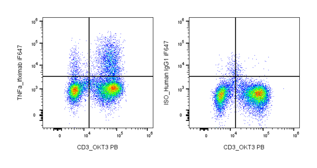
| Clone | Infliximab |
|---|---|
| Application | Flow Cytometry |
| Reactivity | Human |
| Format | iF647 |
| Target Name | TNF-α, TNF-alpha, Tumor necrosis factor-α, Macrophage cytotoxic factor (MCF) |
| Isotype | Human IgG1 |
| Antibody Type | Monoclonal |
| Regulatory Status | RUO |
| Formulation | Phosphate-buffered solution, pH 7.2, containing 0.09% sodium azide and 0.2% (w/v) BSA |
| Protein Concentration | Supplied at a lot-specific concentration. |
| Storage&Handling | The antibody solution should be stored undiluted between 2°C and 8°C, and protected from prolonged exposure to light. Do not freeze. |
| Recommended Usage | For flow cytometric staining, it is recommended to use 5 µL of this reagent per 0.5-1.0 million cells in a 100 µL volume. Optimal reagent performance should be determined by titration for each specific application. iF647 has an excitation max at 656 nm and an emission max at 670 nm. |
| Excitation Laser | Red Laser (633 nm) |
| See All Formats | Clone Infliximab |
TNFα (tumor necrosis factor alpha) is a potent pro-inflammatory cytokine that plays a central role in immune regulation, host defense, and inflammation. It is primarily produced by activated macrophages, T cells, and other immune cells in response to infection, injury, or immune stimulation. TNFα mediates a wide range of biological effects, including induction of fever, activation of endothelial cells, promotion of leukocyte recruitment, and regulation of cell survival, apoptosis, and necrosis.
Structurally, TNFα is initially synthesized as a type II transmembrane protein (membrane-bound TNFα) that forms stable homotrimers. It can be cleaved by the metalloprotease TACE (ADAM17) to release a soluble trimeric form. Both membrane-bound and soluble TNFα are biologically active, although they may have distinct functional roles. The trimeric structure is essential for binding and activating its receptors.
The primary receptors for TNFα are TNFR1 (p55, CD120a) and TNFR2 (p75, CD120b). TNFR1 is widely expressed and contains a death domain that can trigger apoptosis or activate NF-κB signaling pathways, leading to inflammation and cell survival. TNFR2 is more restricted in expression and is mainly involved in immune regulation and tissue repair. The interaction between TNFα and its receptors initiates complex signaling cascades that determine cellular outcomes.
In disease, TNFα is a key driver of chronic inflammatory and autoimmune conditions, including rheumatoid arthritis, inflammatory bowel disease, psoriasis, and ankylosing spondylitis. Excessive or dysregulated TNFα production contributes to tissue damage and disease progression. TNFα is also involved in cancer, infection, and sepsis, where it can have both protective and pathological effects.
Therapeutically, TNFα is one of the most successfully targeted cytokines in modern medicine. Anti-TNF biologics, such as monoclonal antibodies and receptor fusion proteins, have revolutionized the treatment of inflammatory diseases by neutralizing TNFα activity. These therapies reduce inflammation and improve clinical outcomes, although they may increase susceptibility to infections due to immune suppression.
iF647 Human IgG1 Isotype Control Antibody
iF647 Anti-Human TNF-α Antibody TDS
Have a product or application question? Consult our FAQs or contact us.