| Cat # | Size | Price | Quantity | |
|---|---|---|---|---|
| 112201 | 25 tests | $140 | ||
| 112202 | 100 tests | $320 |
| Clone | Tiragolumab |
|---|---|
| Application | Flow Cytometry |
| Reactivity | Human |
| Format | PE |
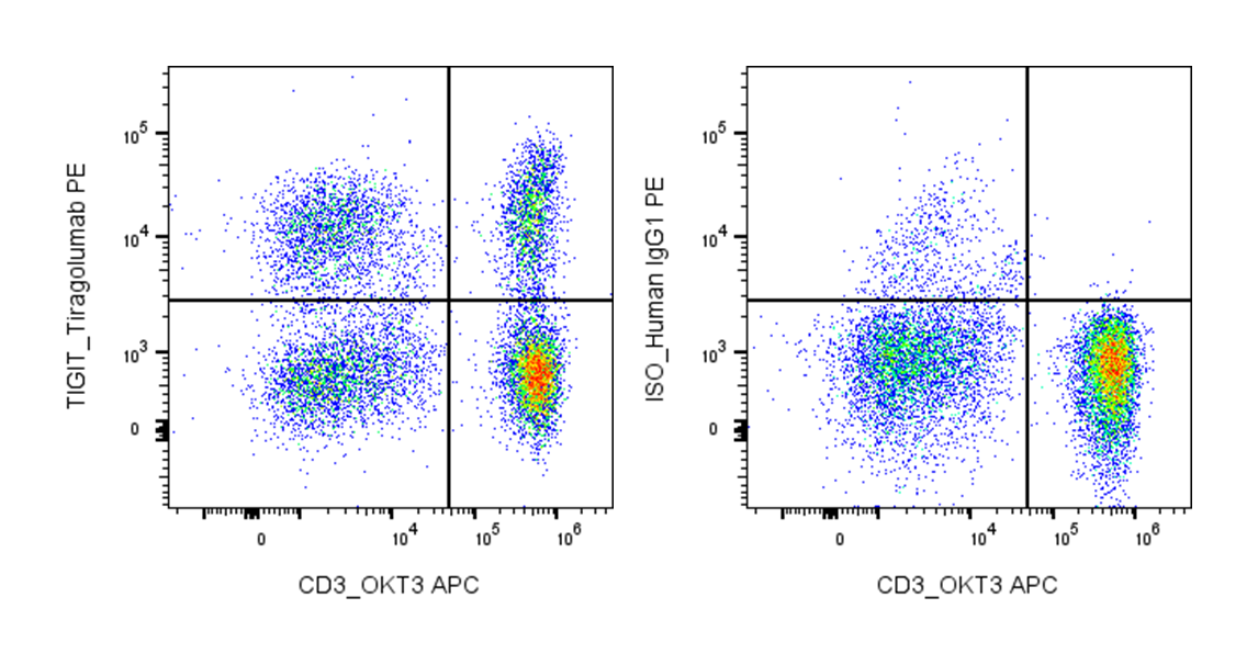
| Target Name | TIGIT, VSTM3, T-cell immunoreceptor with Ig and ITIM domains, VSIG9 |
| Isotype | Human IgG1 |
| Antibody Type | Monoclonal |
| Regulatory Status | RUO |
| Formulation | Phosphate-buffered solution, pH 7.2, containing 0.09% sodium azide and 0.2% (w/v) BSA |
| Protein Concentration | Supplied at a lot-specific concentration. |
| Storage&Handling | The antibody solution should be stored undiluted between 2°C and 8°C, and protected from prolonged exposure to light. Do not freeze. |
| Recommended Usage | For flow cytometric staining, it is recommended to use 5 µL of this reagent per 0.5-1.0 million cells in a 100 µL volume. Optimal reagent performance should be determined by titration for each specific application. PE has an excitation max at 565 nm and an emission max at 575 nm. |
| Excitation Laser | Blue Laser (488 nm) Green/Yellow laser (532/561nm) |
| See All Formats | Clone Tiragolumab |
Human TIGIT (T cell immunoreceptor with Ig and ITIM domains) is an inhibitory receptor expressed on multiple immune cells, including activated T cells, regulatory T cells (Tregs), and natural killer (NK) cells. It plays a key role in maintaining immune homeostasis by dampening immune responses. Upon engagement, TIGIT suppresses T cell activation, reduces cytokine production, and enhances the suppressive function of Tregs, thereby contributing to immune tolerance.
Structurally, TIGIT is a type I transmembrane glycoprotein belonging to the immunoglobulin superfamily. It contains a single extracellular Ig variable (IgV)-like domain responsible for ligand binding, a transmembrane region, and a cytoplasmic tail that includes an immunoreceptor tyrosine-based inhibitory motif (ITIM) and an immunoglobulin tyrosine tail (ITT)-like motif. These intracellular motifs mediate inhibitory signaling through recruitment of phosphatases and downstream signaling molecules that attenuate immune cell activation.
The primary ligands for TIGIT are CD155 (also known as PVR) and CD112 (Nectin-2), which are widely expressed on antigen-presenting cells and many tumor cells. TIGIT competes with the activating receptor CD226 (DNAM-1) for binding to these ligands, thereby shifting the balance toward immune inhibition. This competitive interaction is central to its regulatory function in immune responses.
In disease, TIGIT is often upregulated in chronic infections and cancer, where it contributes to T cell exhaustion and immune evasion. High TIGIT expression has been observed in tumor-infiltrating lymphocytes across multiple cancer types, including lung, colorectal, and melanoma. Its role in suppressing anti-tumor immunity makes it a critical checkpoint in the tumor microenvironment.
Therapeutically, TIGIT has emerged as a promising target in cancer immunotherapy. Monoclonal antibodies that block TIGIT are being developed to restore T cell and NK cell activity, often in combination with PD-1 or PD-L1 inhibitors. Early clinical studies suggest that targeting TIGIT may enhance anti-tumor responses, particularly in patients who do not fully respond to existing checkpoint therapies. Ongoing research continues to evaluate its efficacy and safety across various malignancies.
PE Human IgG1 Isotype Control Antibody
PE Anti-Human TIGIT (VSTM3) Antibody TDS
Have a product or application question? Consult our FAQs or contact us.