| Cat # | Size | Price | Quantity | |
|---|---|---|---|---|
| 101709 | 25 tests | $130 | ||
| 101710 | 100 tests | $300 |
| Clone | EH12.2H7 |
|---|---|
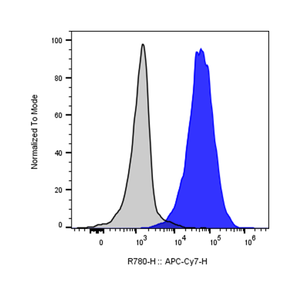
| Application | Flow Cytometry |
| Reactivity | Human |
| Format | APC/Cyanine7 |
| Target Name | CD279, PD1, PD-1 |
| Isotype | Mouse IgG1 |
| Antibody Type | Monoclonal |
| Regulatory Status | RUO |
| Formulation | Phosphate-buffered solution, pH 7.2, containing 0.09% sodium azide and 0.2% (w/v) BSA |
| Protein Concentration | Supplied at a lot-specific concentration. |
| Storage&Handling | The antibody solution should be stored undiluted between 2°C and 8°C, and protected from prolonged exposure to light. Do not freeze. |
| Recommended Usage | For flow cytometric staining, it is recommended to use 5 uL of this reagent per 0.5-1.0 million cells in a 100 µL volume. Optimal reagent performance should be determined by titration for each specific application. APC/Cyanine7 has an excitation max at 650 nm and an emission max at 774 nm. |
| Excitation Laser | Red Laser (633 nm) |
| See All Formats | Clone EH12.2H7 |
CD279, also known as Programmed Cell Death Protein 1 (PD-1), is a crucial immune checkpoint receptor that regulates T cell activation and prevents autoimmunity. This transmembrane protein plays a pivotal role in maintaining immune homeostasis by delivering inhibitory signals that dampen excessive immune responses.
PD-1 is a type I transmembrane glycoprotein belonging to the immunoglobulin superfamily. It contains an extracellular immunoglobulin variable (IgV)-like domain, a transmembrane region, and an intracellular tail with two tyrosine-based signaling motifs: an immunoreceptor tyrosine-based inhibitory motif (ITIM) and an immunoreceptor tyrosine-based switch motif (ITSM). When engaged, these motifs recruit phosphatases that inhibit T-cell receptor signaling, effectively suppressing T-cell activation, proliferation, and cytokine production.
PD-1 interacts with two primary ligands: PD-L1 (B7-H1/CD274) and PD-L2 (B7-DC/CD273). PD-L1 is widely expressed on various cell types, including tumor cells, antigen-presenting cells, and non-hematopoietic tissues, while PD-L2 expression is more restricted to antigen-presenting cells. These ligand-receptor interactions serve as critical brakes on immune responses. In cancer, tumor cells exploit the PD-1/PD-L1 pathway to evade immune surveillance. By upregulating PD-L1 expression, tumors effectively "turn off" infiltrating T-cells, preventing effective anti-tumor immunity. This mechanism contributes to tumor progression and immune escape across multiple cancer types.
The discovery of PD-1's role in cancer has revolutionized oncology through immune checkpoint inhibitors. Monoclonal antibodies targeting PD-1 (pembrolizumab, nivolumab) or PD-L1 (atezolizumab, durvalumab) block this inhibitory pathway, reinvigorating anti-tumor T-cell responses. These therapies have demonstrated remarkable success in treating melanoma, non-small cell lung cancer, renal cell carcinoma, and numerous other malignancies, fundamentally transforming cancer treatment paradigms and offering durable responses in previously untreatable cancers.
APC/Cyanine7 Mouse IgG1 Isotype Control Antibody
APC/Cyanine7 Anti-Human CD279 (PD1) Antibody TDS
PE Human PD-L1 (CD274) Protein (C-His)
Anti-Human PD-L1 (Atezolizumab Biosimilar), Clone Atezolizuma
Anti-Human PD-L1 (Avelumab Biosimilar), Clone Avelumab
Human PD1 (CD279) Protein (C-His-Avi)
Biotin Human PD1 (CD279) Protein (C-His-Avi)
Human PD1 (CD279) Protein (C-His)
Anti-Human PD1 (Camrelizumab Biosimilar), Clone Camrelizumab
Anti-Human PD1 (Cemiplimab Biosimilar), Clone Cemiplimab
Anti-Human PD1 (Nivolumab Biosimilar), Clone Nivolumab
Have a product or application question? Consult our FAQs or contact us.