| Cat # | Size | Price | Quantity | |
|---|---|---|---|---|
| 810501 | 25 ug | $145 | ||
| 810502 | 100 ug | $295 |
| Application | ELISA, BLI |
|---|---|
| Format | Liquid, Purified |
| Expression Host | CHO |
| Target Name | CD3-epsilon, FLJ18683, T3E, TCRE, CD3E |
| Species | Human |
| Sources | Recombinant Human CD3 Protein ( Asp 23- Asp126) with C-terminus Fc-tag is expressed in CHO cell. |
| Accession Number | P07766 |
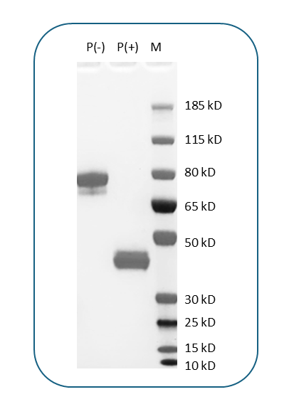
| Molecular Weight | The protein has a predicted molecular weight of 37.9 kDa. Under DTT-reducing conditions, it migrates at approximately 45 kDa on SDS-PAGE. |
| Affinity Tag | C-Fc |
| Purity | >95% based on SDS-PAGE under reducing condition |
| Regulatory Status | RUO |
| Formulation | 1xPBS buffer, pH7.4, 0.22 µm filtered |
| Endotoxin level | Less than 0.1 EU/µg protein as determined by the LAL method |
| Protein Concentration | 25µg size is bottled at 0.2mg/mL concentration. 100 µg size is supplied at a lot-specific concentration. |
| Storage and Handling | Briefly centrifuge the vial upon receipt. An unopened vial can be stored at 4°C for up to 2 weeks, or at -20°C or below for up to six months. The protein may be further diluted to 0.1 mg/mL using 0.22 µm-filtered PBS buffer (pH 7.4). For long-term storage, the diluted stock solution should be aliquoted and stored at ≤ –70°C to minimize freeze-thaw cycles. If additional dilution is required, carrier proteins such as FBS or BSA should be added to maintain protein stability. |
CD3ε (CD3 epsilon) is a critical component of the T cell receptor (TCR) complex and plays an essential role in T cell activation and adaptive immune responses. CD3ε is one of four CD3 chains (γ, δ, ε, and ζ) that associate non-covalently with the TCR α and β chains to form the complete TCR-CD3 complex on the surface of T lymphocytes. While the TCR chains recognize specific antigens presented by major histocompatibility complex (MHC) molecules, the CD3 chains, including CD3ε, are responsible for signal transduction. CD3ε transmits activation signals into the cell following antigen recognition, initiating a cascade of intracellular events that lead to T cell proliferation, differentiation, and effector functions such as cytokine production and cytotoxic activity. The protein is essential for T cell development and the proper assembly and surface expression of the TCR-CD3 complex.
Structurally, CD3ε is a type I transmembrane glycoprotein of approximately 20 kDa belonging to the immunoglobulin superfamily. The extracellular domain contains a single immunoglobulin-like fold that participates in the assembly and stabilization of the TCR-CD3 complex. CD3ε forms heterodimers with either CD3γ or CD3δ chains through non-covalent interactions in the extracellular region. The transmembrane domain contains charged residues that interact with the TCR chains to ensure proper complex assembly. Most importantly, the cytoplasmic tail of CD3ε contains one immunoreceptor tyrosine-based activation motif (ITAM), a conserved signaling sequence that becomes phosphorylated upon TCR engagement. The CD3 epsilon immune recognition receptor cytoplasmic domain also binds to acidic and mixed phospholipid vesicles with a binding strength that correlates with membrane composition. When phosphorylated, the ITAM recruits kinases such as ZAP-70, initiating downstream signaling pathways including the MAPK, PI3K/AKT, and NF-κB cascades that drive T cell activation.
CD3ε does not bind traditional extracellular ligands directly. Instead, its function is triggered when the associated TCR recognizes peptide-MHC complexes on antigen-presenting cells. This recognition event induces conformational changes in the TCR-CD3 complex that expose the cytoplasmic ITAMs for phosphorylation. CD3ε works in concert with the other CD3 chains to amplify and sustain TCR signaling, ensuring robust T cell responses to antigen stimulation and playing a crucial role in T cell development and the initiation of the TCR-CD3 complex assembly.
In disease contexts, mutations or deficiencies in CD3ε can cause severe combined immunodeficiency (SCID), characterized by absent or dysfunctional T cells and profound susceptibility to infections. Conversely, aberrant CD3 signaling contributes to autoimmune diseases and T cell malignancies. CD3ε has also been identified as a prognostic marker in several cancers, including breast invasive carcinoma, cervical squamous cell carcinoma and endocervical adenocarcinoma, and head and neck squamous cell carcinoma. Therapeutically, CD3ε has become one of the most important targets in immunotherapy. Anti-CD3ε antibodies such as muromonab-CD3 (OKT3) were among the first monoclonal antibodies used clinically for immunosuppression in transplant rejection. More recently, bispecific T cell engagers (BiTEs) that bind both CD3ε and tumor-associated antigens redirect T cells to kill cancer cells. Blinatumomab, a CD3ε/CD19 BiTE, is approved for acute lymphoblastic leukemia. Additionally, CD3ε-targeting antibodies are being developed for autoimmune diseases and as components of chimeric antigen receptor (CAR) constructs, establishing CD3ε as a cornerstone target in modern immunotherapy across oncology, transplantation, and autoimmunity.
Anti-Human CD4 Antibody, Clone 004AB
iF488 Anti-Human CD4 Antibody, Clone OKT4-Rec
iF647 Anti-Human CD4 Antibody, Clone OKT4-Rec
PE Anti-Human CD4 Antibody, Clone RPA-T4
Anti-Human CD3 Antibody, Clone Hit3a
iF700 Anti-Human CD3 Antibody, Clone Hit3a
iF488 Anti-Human CD3 Antibody, Clone OKT3
PerCP/Cyanine5.5 Anti-Human CD3 Antibody, Clone OKT3
PE Anti-Human CD3 Antibody, Clone SP34
iF647 Anti-Human CD3 Antibody, Clone SP34
Have a product or application question? Consult our FAQs or contact us.