| Cat # | Size | Price | Quantity | |
|---|---|---|---|---|
| 805401 | 25 ug | $145 | ||
| 805402 | 100 ug | $295 |
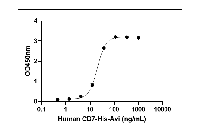
| Application | ELISA, BLI |
|---|---|
| Format | Liquid, Purified |
| Expression Host | CHO |
| Target Name | CD7, GP40, TP41, LEU-9, Tp40 |
| Species | Human |
| Sources | Recombinant Human CD7 (Ala26-Pro180) with C-terminus His tag & Avi tag is expressed in CHO cell |
| Accession Number | P09564 |
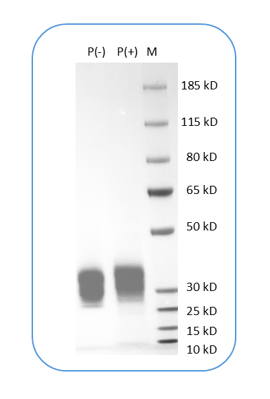
| Molecular Weight | The protein has a predicted molecular weight of 18 kDa. Under DTT-reducing conditions, it migrates at approximately 30-40 kDa on SDS-PAGE. |
| Affinity Tag | C-His-Avi |
| Purity | >95% based on SDS-PAGE under reducing condition |
| Regulatory Status | RUO |
| Formulation | 1xPBS buffer, pH7.4, 0.22 µm filtered |
| Endotoxin level | Not tested |
| Protein Concentration | 25µg size is bottled at 0.2mg/mL concentration. 100 µg size is supplied at a lot-specific concentration. |
| Storage and Handling | Briefly centrifuge the vial upon receipt. An unopened vial can be stored at 4°C for up to 2 weeks, or at -20°C or below for up to six months. The protein may be further diluted to 0.1 mg/mL using 0.22 µm-filtered PBS buffer (pH 7.4). For long-term storage, the diluted stock solution should be aliquoted and stored at ≤ –70°C to minimize freeze-thaw cycles. If additional dilution is required, carrier proteins such as FBS or BSA should be added to maintain protein stability. |
CD7 is a transmembrane glycoprotein belonging to the immunoglobulin superfamily that serves as an important marker of T cell and natural killer (NK) cell lineages. It is one of the earliest surface antigens expressed during T cell development, appearing on thymocytes and persisting throughout T cell maturation. CD7 plays a costimulatory role in T cell activation and is involved in regulating immune responses. The protein participates in T cell receptor signaling, cell-cell interactions, and may contribute to immune synapse formation and lymphocyte trafficking, though its complete physiological functions continue to be investigated.
Structurally, CD7 is a type I transmembrane protein of approximately 40 kDa consisting of a single extracellular immunoglobulin-like domain, a transmembrane region, and a cytoplasmic tail. The extracellular domain contains the characteristic features of the immunoglobulin superfamily, including disulfide bonds that stabilize its three-dimensional structure. The cytoplasmic domain is relatively short but contains motifs that can interact with intracellular signaling molecules and cytoskeletal components. CD7 can form homodimers on the cell surface, and this oligomerization may be important for its signaling and costimulatory functions.
The primary identified ligand for CD7 is K12/SECTM1 (secreted and transmembrane 1), which binds to CD7 and mediates costimulatory signals during T cell activation. Upon binding to K12/SECTM1, CD7 triggers the production of cytokines and enhances T cell responses. Additionally, CD7 may engage in homophilic interactions (CD7-CD7 binding between cells) and has been reported to interact with galectin-1, a lectin involved in immune regulation. The protein's glycosylation patterns influence its binding properties and functional activities in the immune system.
In disease contexts, CD7 expression patterns are clinically significant in hematologic malignancies. CD7 is expressed on most T cell acute lymphoblastic leukemias (T-ALL) and certain peripheral T cell lymphomas, making it a valuable diagnostic marker. CD7-positive acute myeloid leukemia (AML) represents a subset with distinct clinical features and often poorer prognosis. Therapeutically, CD7 has emerged as a promising target for immunotherapy in T cell malignancies. CD7-directed chimeric antigen receptor (CAR) T cell therapies are under active development and clinical investigation for treating T-ALL and peripheral T cell lymphomas. However, targeting CD7 presents unique challenges because normal T cells also express this marker, requiring innovative strategies such as CD7 knockout in CAR-T cells to prevent fratricide (self-destruction). K12/SECTM1-based CAR T cells that exploit the natural CD7-K12 interaction are being developed to enhance specificity and efficacy while minimizing toxicity to normal T cells.
Human CD7 Protein (C-His-Avi) TDS
Biotin Human CD7 Protein (C-His-Avi)
Have a product or application question? Consult our FAQs or contact us.