| Cat # | Size | Price | Quantity | |
|---|---|---|---|---|
| 106805 | 25 tests | $90 | ||
| 106806 | 100 tests | $210 |
| Clone | p282 (H19) |
|---|---|
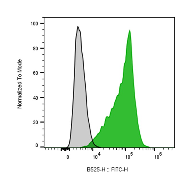
| Application | Flow Cytometry |
| Reactivity | Human |
| Format | iF488 |
| Target Name | CD59, Protectin, MIRL, H19, MACIF |
| Isotype | Mouse IgG2a |
| Antibody Type | Monoclonal |
| Regulatory Status | RUO |
| Formulation | Phosphate-buffered solution, pH 7.2, containing 0.09% sodium azide and 0.2% (w/v) BSA |
| Protein Concentration | Supplied at a lot-specific concentration. |
| Storage&Handling | The antibody solution should be stored undiluted between 2°C and 8°C, and protected from prolonged exposure to light. Do not freeze. |
| Recommended Usage | For flow cytometric staining, it is recommended to use 5 uL of this reagent per 0.5-1.0 million cells in a 100 µL volume. Optimal reagent performance should be determined by titration for each specific application. |
| Excitation Laser | Blue Laser (488 nm) |
| See All Formats | Clone p282 (H19) |
CD59, also known as protectin, is a glycosylphosphatidylinositol (GPI)-anchored cell surface protein that plays a crucial role in regulating the complement system. It is widely expressed on hematopoietic cells, endothelial cells, epithelial cells, and many other tissues, where it protects host cells from unintended complement-mediated damage.
Structurally, CD59 is a small (~18–20 kDa) glycoprotein composed of a single extracellular domain stabilized by disulfide bonds and attached to the plasma membrane via a GPI anchor. Unlike transmembrane receptors, CD59 lacks an intracellular signaling domain and functions primarily through extracellular interactions. Its structure is optimized to bind late complement components and interfere with assembly of the terminal complement complex. Functionally, CD59 acts as a potent inhibitor of the membrane attack complex (MAC), the lytic pore formed by complement components C5b, C6, C7, C8, and multiple copies of C9. CD59 specifically binds to C8 and C9, preventing C9 polymerization and insertion into the cell membrane. By blocking MAC formation, CD59 protects self cells from complement-mediated lysis during normal immune responses and inflammatory conditions. This regulatory role is essential for maintaining tissue integrity while allowing complement to target pathogens.
CD59 is implicated in several diseases when its expression or function is altered. The most prominent example is paroxysmal nocturnal hemoglobinuria (PNH), a rare hematologic disorder caused by acquired mutations affecting GPI anchor biosynthesis. In PNH, red blood cells lack CD59 (and other GPI-anchored proteins), rendering them highly susceptible to complement-mediated destruction and leading to chronic hemolytic anemia and thrombosis. Reduced or dysregulated CD59 expression has also been observed in autoimmune diseases, neuroinflammatory disorders, and ischemia-reperfusion injury, where insufficient complement regulation contributes to tissue damage. Conversely, many tumors upregulate CD59 to protect themselves from complement-dependent cytotoxicity.
Therapeutically, CD59 is relevant in both complement-targeted treatments and cancer immunotherapy. In PNH and related complement-mediated diseases, therapies such as C5 inhibitors indirectly compensate for the loss of CD59 by preventing MAC formation. In oncology, strategies to block or downregulate CD59 on tumor cells are being explored to enhance the efficacy of antibody-based therapies that rely on complement-dependent cytotoxicity. Additionally, CD59 serves as a useful biomarker in flow cytometry for diagnosing PNH and assessing complement regulatory status, highlighting its importance in both clinical diagnostics and therapeutic development.
iF488 Mouse IgG2a Isotype Control Antibody
iF488 Anti-Human CD59 Antibody TDS
Have a product or application question? Consult our FAQs or contact us.