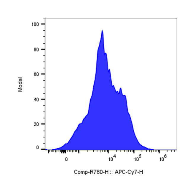
| Cat # | Size | Price | Quantity | |
|---|---|---|---|---|
| 200509 | 25 tests | $95 | ||
| 200510 | 100 tests | $200 |
| Clone | MEL-14 |
|---|---|
| Application | Flow Cytometry |
| Reactivity | Mouse |
| Format | APC/Cyanine7 |
| Target Name | CD62L, LAM-1, L-selectin, Ly-22 |
| Isotype | Rat IgG2a |
| Antibody Type | Monoclonal |
| Regulatory Status | RUO |
| Formulation | Phosphate-buffered solution, pH 7.2, containing 0.09% sodium azide and 0.2% (w/v) BSA |
| Protein Concentration | Supplied at a lot-specific concentration. |
| Storage&Handling | The antibody solution should be stored undiluted between 2°C and 8°C, and protected from prolonged exposure to light. Do not freeze. |
| Recommended Usage | For flow cytometric staining, it is recommended to use 5 uL of this reagent per 0.5-1.0 million cells in a 100 µL volume. Optimal reagent performance should be determined by titration for each specific application. |
| Excitation Laser | Red Laser (633 nm) |
| Research Areas | Adhesion Molecules, T cells, Central Memory T cells, B cells, Monocytes, Neutrophils, Eosinophils, NK cells. |
| See All Formats | Clone MEL-14 |
CD62L, also known as L-selectin, is a cell surface adhesion molecule expressed predominantly on most leukocytes, including naïve T cells, B cells, monocytes, neutrophils, and subsets of natural killer cells. It plays an essential role in mediating lymphocyte homing to peripheral lymphoid organs and enables leukocyte recruitment to sites of inflammation. By facilitating the initial “rolling” interactions of immune cells along the vascular endothelium, CD62L helps coordinate immune surveillance and the body’s response to infection or tissue injury.
Structurally, CD62L is a type I transmembrane glycoprotein belonging to the selectin family, which also includes E-selectin and P-selectin. It consists of an N-terminal lectin domain responsible for binding carbohydrate ligands, an epidermal growth factor-like domain, a series of consensus repeats, a single transmembrane region, and a short cytoplasmic tail. The receptor’s extracellular domains mediate specific recognition of sialylated and fucosylated carbohydrate ligands expressed on endothelial cells, allowing for rapid adhesion under shear flow conditions. CD62L can be proteolytically cleaved from the cell surface, generating a soluble form whose levels increase during inflammation.
The principal ligands of CD62L include peripheral node addressins (PNAd) on high endothelial venules within lymph nodes, as well as glycoproteins such as GlyCAM-1, CD34 (in the vascular sense, not the hematopoietic stem cell marker function), and MAdCAM-1. These interactions are essential for naïve and central memory lymphocytes to enter secondary lymphoid tissues, where immune activation and antigen recognition primarily occur.
In disease, dysregulation of CD62L expression or shedding contributes to immune dysfunction. Reduced CD62L expression on T cells is linked with chronic inflammatory and autoimmune diseases, including rheumatoid arthritis and multiple sclerosis, reflecting altered trafficking and immune activation. In cancer, CD62L expression serves as a marker distinguishing central memory from effector T cells and influences antitumor immune responses. Low CD62L on adoptively transferred T cells correlates with diminished persistence and tumor control in immunotherapy models.
Therapeutically, CD62L holds importance in immune cell-based treatments and diagnostics. Monitoring its expression helps identify functional subsets of T cells for adoptive cell therapy and evaluate immune activation states. Modulation of CD62L-ligand interactions offers potential for controlling leukocyte recruitment in inflammatory and autoimmune diseases. Consequently, CD62L remains a key molecular target in efforts to regulate immune cell trafficking and improve immunotherapeutic precision.
APC/Cyanine7 Anti-Mouse CD62L (L-selectin) Antibody TDS
Have a product or application question? Consult our FAQs or contact us.