| Cat # | Size | Price | Quantity | |
|---|---|---|---|---|
| 120401 | 25 µg | $45 | ||
| 120402 | 100 µg | $95 |
| Clone | 4G7 |
|---|---|
| Application | Flow Cytometry |
| Reactivity | Human |
| Format | Purified |
| Target Name | CD19, B4, CVID3, MGC12802 |
| Isotype | mouse IgG1 |
| Antibody Type | Monoclonal |
| Regulatory Status | RUO |
| Formulation | Phosphate-buffered solution, pH 7.2, containing 0.09% sodium azide |
| Protein Concentration | 0.5 mg/mL |
| Storage & Handling | The antibody solution should be stored between 2°C and 8°C |
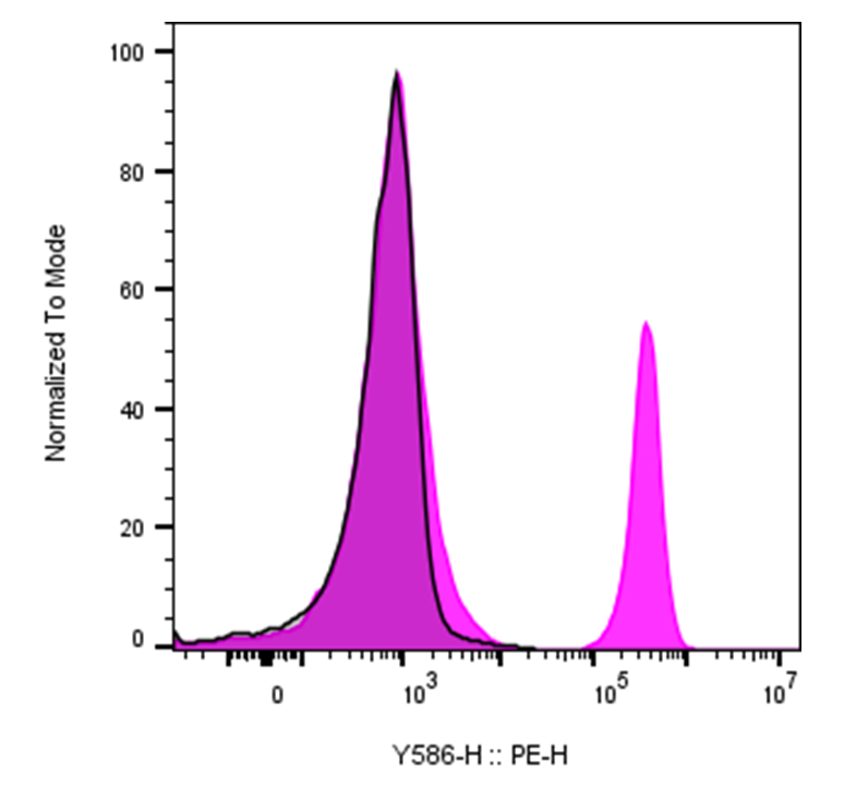
| Recommended Usage | For flow cytometric staining, it is recommended to use less than 0.2 µg of this reagent per 0.5-1.0 million cells in a 100 µL volume. Optimal reagent performance should be determined by titration for each specific application |
| See All Formats | Clone 4G7 |
CD19 is a B cell-specific transmembrane glycoprotein that serves as a central regulator of B cell activation, signaling, and development. It is expressed throughout most stages of B cell differentiation, from early pro-B cells through mature peripheral B cells, but is lost upon terminal differentiation into plasma cells. Because of its broad and consistent expression on B lineage cells, CD19 is widely used as a defining marker of the B cell compartment.
Structurally, CD19 is a type I transmembrane protein with two extracellular immunoglobulin-like domains, a single transmembrane region, and a long cytoplasmic tail. The intracellular domain contains multiple tyrosine residues that become phosphorylated following B cell receptor (BCR) engagement. CD19 functions as part of a larger co-receptor complex that includes CD21 (complement receptor 2), CD81 (TAPA-1), and CD225, which together modulate BCR signaling strength and sensitivity.
Functionally, CD19 acts as a signaling amplifier for the BCR. When antigen binds the BCR, CD19 is rapidly phosphorylated by Src family kinases, creating docking sites for signaling molecules such as PI3K. This lowers the threshold for B cell activation, enhances proliferation, promotes survival, and supports antibody production. Through this role, CD19 helps shape humoral immune responses and ensures efficient activation of B cells in response to antigen, particularly when antigen is present at low concentrations. Unlike many immune receptors, CD19 does not have a well-defined classical ligand. Instead, its activity is regulated through its association with the BCR complex and co-receptors. The CD19-CD21 interaction is functionally linked to recognition of complement-tagged antigens, indirectly coupling innate and adaptive immune signals to enhance B cell responses.
Dysregulation of CD19 signaling contributes to disease. Overactive CD19-mediated signaling has been associated with autoimmune diseases such as systemic lupus erythematosus, where hyperresponsive B cells drive autoantibody production. CD19 is also expressed on the vast majority of B cell malignancies, including B cell acute lymphoblastic leukemia (B-ALL), chronic lymphocytic leukemia, and many non-Hodgkin lymphomas, making it a critical diagnostic and therapeutic target.
In therapeutics, CD19 is one of the most successful targets in modern immuno-oncology. CD19-directed therapies include monoclonal antibodies, antibody–drug conjugates, bispecific T cell engagers, and chimeric antigen receptor (CAR) T cell therapies, many of which have produced durable remissions in refractory B cell cancers. Beyond cancer, CD19-targeted strategies are being explored to selectively deplete pathogenic B cells in autoimmune disease, underscoring CD19’s central role in both disease biology and therapeutic innovation.
Mouse IgG1 Isotype Control Antibody
Have a product or application question? Consult our FAQs or contact us.