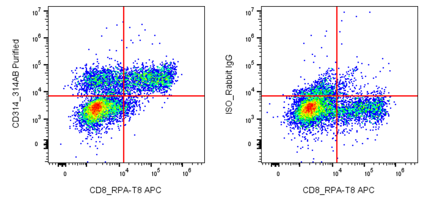
| Cat # | Size | Price | Quantity | |
|---|---|---|---|---|
| 121101 | 25 µg | $55 | ||
| 121102 | 100 µg | $125 |
| Clone | 314AB |
|---|---|
| Application | Flow Cytometry |
| Reactivity | Human |
| Format | Purified |
| Target Name | CD314, NKG2D |
| Isotype | Rabbit IgG |
| Antibody Type | Monoclonal |
| Regulatory Status | RUO |
| Formulation | Phosphate-buffered solution, pH 7.2, containing 0.09% sodium azide |
| Protein Concentration | 0.5 mg/mL |
| Storage & Handling | The antibody solution should be stored between 2°C and 8°C |
| Recommended Usage | For flow cytometric staining, it is recommended to use less than 0.2 µg of this reagent per 0.5-1.0 million cells in a 100 µL volume. Optimal reagent performance should be determined by titration for each specific application |
| See All Formats | Clone 314AB |
CD314, also known as NKG2D and encoded by the KLRK1 gene, is an activating immune receptor expressed primarily on natural killer (NK) cells and on subsets of CD8+ T cells and other cytotoxic lymphocytes. NKG2D plays an important role in immune surveillance by recognizing stress-induced molecules that are frequently upregulated on infected, transformed, or otherwise damaged cells. Engagement of CD314 promotes cytotoxicity, cytokine production, and other effector functions, allowing NK and T cells to eliminate abnormal cells.
CD314 is a type II transmembrane C-type lectin-like receptor that contains a short cytoplasmic region, a transmembrane domain, and an extracellular C-type lectin-like domain. Unlike many activating receptors, NKG2D does not signal through its own long cytoplasmic tail. Instead, its transmembrane region associates with the adaptor protein DAP10, which contains a signaling motif that activates PI3K and other downstream pathways. In humans, the major ligands for CD314 are the stress-induced MHC class I-related molecules MICA and MICB and members of the UL16-binding protein family, including ULBP1–6.
The CD314 ligand system is important in cancer, viral infection, and immune-mediated disease. Tumor cells and infected cells can increase expression of NKG2D ligands, making them targets for NK- and T-cell-mediated killing. However, chronic ligand expression or persistent immune stimulation may contribute to NKG2D dysfunction and immune exhaustion. Tumors may also evade immune surveillance by releasing soluble forms of MICA, MICB, or other NKG2D ligands, potentially reducing effective receptor signaling.
Because CD314 is a potent activator of cytotoxic lymphocytes, the NKG2D pathway is being explored for cancer immunotherapy. Strategies include NKG2D-based CAR-T and CAR-NK cells designed to recognize multiple stress-associated ligands, as well as approaches that enhance NKG2D signaling or prevent tumor-mediated ligand shedding. These strategies aim to improve recognition and elimination of malignant cells while addressing the challenges associated with ligand heterogeneity and immune escape.
Rabbit IgG, κ2 Isotype Control Antibody
Anti-human CD314 (NKG2D) Antibody TDS
Have a product or application question? Consult our FAQs or contact us.