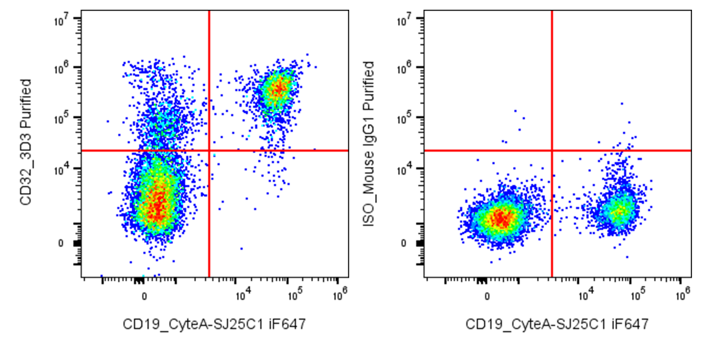
| Cat # | Size | Price | Quantity | |
|---|---|---|---|---|
| 120601 | 25 µg | $75 | ||
| 120602 | 100 µg | $175 |
| Clone | 3D3 |
|---|---|
| Application | Flow Cytometry |
| Reactivity | Human |
| Format | Purified |
| Target Name | CD32, FcγRII, CD32a/FcγRIIa/FCGR2A, CD32b/FcγRIIb/FCGR2B |
| Isotype | mouse IgG1 |
| Antibody Type | Monoclonal |
| Regulatory Status | RUO |
| Formulation | Phosphate-buffered solution, pH 7.2, containing 0.09% sodium azide |
| Protein Concentration | 0.5 mg/mL |
| Storage & Handling | The antibody solution should be stored between 2°C and 8°C |
| Recommended Usage | For flow cytometric staining, it is recommended to use less than 0.2 µg of this reagent per 0.5-1.0 million cells in a 100 µL volume. Optimal reagent performance should be determined by titration for each specific application |
| See All Formats | Clone 3D3 |
Human CD32, also known as Fcγ receptor II (FcγRII), is a family of low-affinity receptors for the Fc portion of immunoglobulin G (IgG) that links antibody-mediated recognition to cellular immune responses. The family comprises three closely related receptors, CD32A (FcγRIIA), CD32B (FcγRIIB), and CD32C (FcγRIIC). They are expressed on various immune cells, including monocytes, macrophages, neutrophils, dendritic cells, and B cells, with expression patterns differing among the isoforms. :contentReference[oaicite:0]{index=0}
CD32 proteins are type I transmembrane glycoproteins belonging to the immunoglobulin superfamily. Their extracellular region contains two Ig-like domains that bind the Fc region of IgG, followed by a transmembrane segment and a cytoplasmic signaling region. CD32A and CD32C contain immunoreceptor tyrosine-based activation motifs (ITAMs), whereas CD32B contains an immunoreceptor tyrosine-based inhibitory motif (ITIM). CD32 has relatively low affinity for monomeric IgG but efficiently binds multivalent IgG-containing immune complexes. :contentReference[oaicite:1]{index=1}
The principal ligands are IgG antibodies and IgG-containing immune complexes, with receptor affinity varying according to IgG subclass and CD32 isoform. CD32A and CD32C generally promote cellular activation, phagocytosis, cytokine production, and inflammatory responses, while CD32B counterbalances these activities and helps maintain immune tolerance. :contentReference[oaicite:2]{index=2}
Dysregulated CD32 signaling is implicated in autoimmune and inflammatory diseases, infectious disease, and cancer. In particular, CD32A genetic variants have been associated with susceptibility to several immune-mediated diseases, while CD32B contributes to regulation of B-cell and myeloid-cell activation. Therapeutically, CD32 is important both as a mediator of monoclonal-antibody activity and as a potential target itself. CD32B-directed antibodies have demonstrated antitumor activity in preclinical B-cell lymphoma models, while soluble FcγR-based approaches and CD32A antagonists are being investigated for antibody-driven autoimmune and inflammatory diseases. :contentReference[oaicite:3]{index=3}
Mouse IgG1 Isotype Control Antibody
Have a product or application question? Consult our FAQs or contact us.