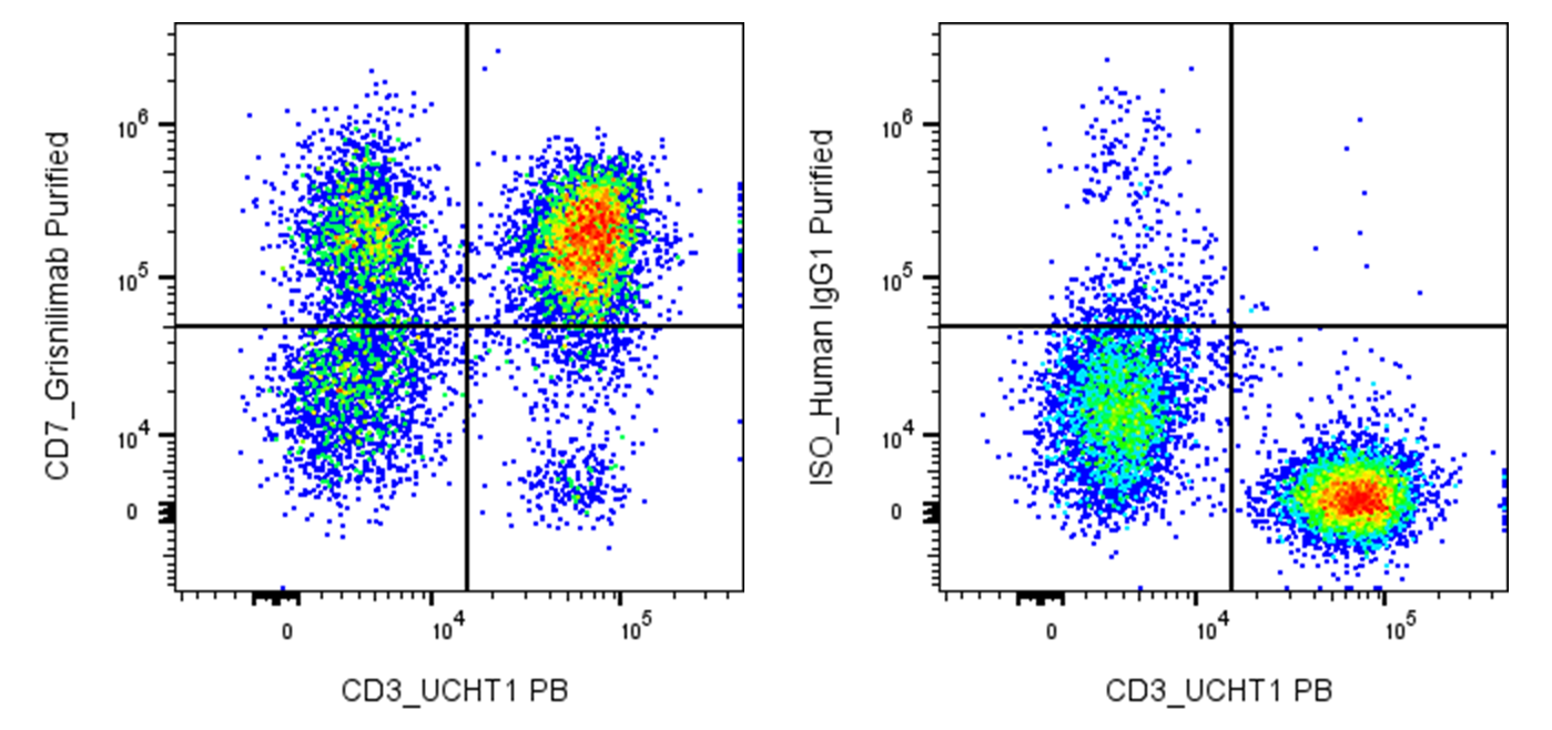
| Cat # | Size | Price | Quantity | |
|---|---|---|---|---|
| 118801 | 25 µg | $35 | ||
| 118802 | 100 µg | $50 |
| Clone | Grisnilimab |
|---|---|
| Application | Flow Cytometry |
| Reactivity | Human |
| Format | Purified |
| Target Name | CD7, gp40 |
| Isotype | Human IgG1 |
| Antibody Type | Monoclonal |
| Regulatory Status | RUO |
| Formulation | Phosphate-buffered solution, pH 7.2, containing 0.09% sodium azide |
| Protein Concentration | 0.5 mg/mL |
| Storage & Handling | The antibody solution should be stored between 2°C and 8°C |
| Recommended Usage | For flow cytometric staining, it is recommended to use less than 0.2 µg of this reagent per 0.5-1.0 million cells in a 100 µL volume. Optimal reagent performance should be determined by titration for each specific application |
| See All Formats | Clone Grisnilimab |
Human CD7 is a 40-kDa type I transmembrane glycoprotein and one of the earliest surface markers expressed during T-cell development. It is present on the majority of mature T cells and natural killer (NK) cells, as well as on early lymphoid and some myeloid progenitors. CD7 belongs to the immunoglobulin superfamily and contains a single extracellular immunoglobulin-like domain, a transmembrane region, and a short cytoplasmic tail containing signaling motifs. CD7 contributes to T-cell and NK-cell activation, adhesion, and cytokine production, functioning primarily as a costimulatory molecule that can amplify signals generated through the T-cell receptor.
The best-characterized ligand for human CD7 is secreted and transmembrane protein 1 (SECTM1), which exists in both membrane-bound and soluble forms. Interaction between SECTM1 and CD7 can promote T-cell proliferation and enhance cytokine production, including interferon-γ and IL-2, particularly in cooperation with other costimulatory signals.
CD7 itself is not generally considered a disease-causing molecule, but its expression pattern makes it an important biomarker and therapeutic target in hematological malignancies. CD7 is expressed in more than 95% of T-cell acute lymphoblastic leukemia (T-ALL) and T-cell lymphoblastic lymphoma (T-LBL), and is also present in subsets of peripheral T-cell lymphomas and acute myeloid leukemia.
Therapeutically, CD7 is being extensively investigated as a target for monoclonal antibodies, antibody-drug conjugates, immunotoxins, and particularly CAR-T-cell therapy. CD7 CAR-T cells have shown promising responses in relapsed or refractory T-ALL/LBL and other CD7-positive malignancies. A major challenge is “fratricide,” because normal T cells and the engineered CAR-T cells themselves express CD7. Gene editing, protein masking, and other engineering strategies are therefore being developed to prevent CAR-T self-killing and improve treatment durability.
Human IgG1 Isotype Control Antibody
Have a product or application question? Consult our FAQs or contact us.