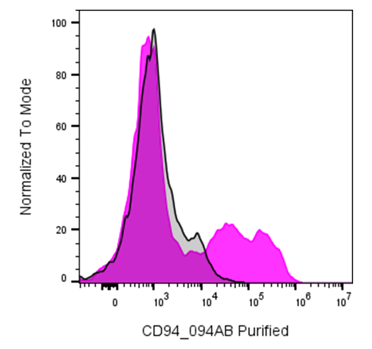
| Cat # | Size | Price | Quantity | |
|---|---|---|---|---|
| 120901 | 25 µg | $55 | ||
| 120902 | 100 µg | $125 |
| Clone | 094AB |
|---|---|
| Application | Flow Cytometry |
| Reactivity | Human |
| Format | Purified |
| Target Name | CD94, KP43 |
| Isotype | Rabbit IgG |
| Antibody Type | Monoclonal |
| Regulatory Status | RUO |
| Formulation | Phosphate-buffered solution, pH 7.2, containing 0.09% sodium azide |
| Protein Concentration | 0.5 mg/mL |
| Storage & Handling | The antibody solution should be stored between 2°C and 8°C |
| Recommended Usage | For flow cytometric staining, it is recommended to use less than 0.2 µg of this reagent per 0.5-1.0 million cells in a 100 µL volume. Optimal reagent performance should be determined by titration for each specific application |
| See All Formats | Clone 094AB |
CD94, encoded by the KLRD1 gene, is a C-type lectin-like receptor expressed primarily on natural killer (NK) cells and subsets of T cells, particularly NKT-like and CD8+ T cells. CD94 plays an important role in regulating cytotoxic lymphocyte activity by recognizing major histocompatibility complex class I (MHC-I)-related molecules and helping NK cells distinguish healthy cells from cells undergoing infection or transformation. Its function depends strongly on the receptor it associates with, allowing CD94-containing complexes to deliver either inhibitory or activating signals.
CD94 is a type II transmembrane glycoprotein belonging to the C-type lectin-like receptor family. It contains a short cytoplasmic region, a transmembrane domain, and an extracellular C-type lectin-like domain. CD94 commonly forms heterodimers with members of the NKG2 family, including NKG2A, NKG2B, NKG2C, and NKG2E. The principal ligands for CD94/NKG2 receptors are HLA-E molecules presenting specific peptides derived largely from the leader sequences of other HLA class I proteins. CD94/NKG2A generally mediates inhibitory signaling, whereas CD94/NKG2C is associated with activating responses through the adaptor protein DAP12.
The CD94/NKG2-HLA-E axis contributes to immune surveillance and is involved in the regulation of antiviral and antitumor immunity. Altered expression or signaling of CD94-containing receptors has been reported in cancer, viral infections, and autoimmune or inflammatory conditions. Tumors may exploit inhibitory CD94/NKG2A signaling to evade NK- and T-cell-mediated immune responses, while certain chronic viral infections can alter the balance of CD94 receptor expression and activity.
Because CD94-containing receptors regulate cytotoxic lymphocyte responses, they are being investigated as potential targets for immunotherapy. Blocking inhibitory NKG2A/CD94 signaling may enhance NK- and T-cell activity against tumors, particularly when combined with other immune checkpoint inhibitors. Conversely, activating pathways involving CD94/NKG2C may potentially enhance antitumor or antiviral immunity. CD94 is therefore a useful marker for studying NK-cell biology and an emerging target for therapeutic strategies aimed at modulating innate and adaptive immune responses.
Rabbit IgG, κ2 Isotype Control Antibody
Have a product or application question? Consult our FAQs or contact us.