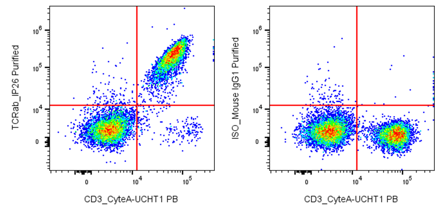
| Cat # | Size | Price | Quantity | |
|---|---|---|---|---|
| 119801 | 25 µg | $50 | ||
| 119802 | 100 µg | $90 |
| Clone | IP26 |
|---|---|
| Application | Flow Cytometry |
| Reactivity | Human |
| Format | Purified |
| Target Name | TCR α/β, α/β TCR |
| Isotype | mouse IgG1 |
| Antibody Type | Monoclonal |
| Regulatory Status | RUO |
| Formulation | Phosphate-buffered solution, pH 7.2, containing 0.09% sodium azide |
| Protein Concentration | 0.5 mg/mL |
| Storage & Handling | The antibody solution should be stored between 2°C and 8°C |
| Recommended Usage | For flow cytometric staining, it is recommended to use less than 0.2 µg of this reagent per 0.5-1.0 million cells in a 100 µL volume. Optimal reagent performance should be determined by titration for each specific application |
| See All Formats | Clone IP26 |
Human T-cell receptor α/β (TCRαβ) is the antigen-recognition receptor expressed by the majority of mature T cells and is central to adaptive cellular immunity. It enables T cells to distinguish foreign, altered-self, and self antigens and, following appropriate recognition, initiates signaling that regulates T-cell activation, proliferation, cytokine production, and cytotoxicity. Unlike antibodies, TCRαβ generally recognizes antigen only when it is presented by another molecule, most commonly a peptide bound to a major histocompatibility complex (MHC).
TCRαβ is a clonotypic heterodimer consisting of one TCRα chain and one TCRβ chain. Each chain contains an extracellular variable (V) and constant (C) immunoglobulin-like domain, a connecting peptide, a transmembrane region, and a short cytoplasmic tail. The variable domains contain three complementarity-determining regions (CDR1, CDR2, and CDR3); the highly diverse CDR3 regions make major contributions to peptide specificity. TCRαβ associates noncovalently with the CD3γε, CD3δε, and CD3ζζ signaling modules, which transmit activation signals after ligand engagement.
The principal ligands for TCRαβ are peptide–MHC class I (pMHC-I) complexes recognized predominantly by CD8 T cells and peptide–MHC class II (pMHC-II) complexes recognized predominantly by CD4 T cells. TCRs can also recognize antigens presented by certain nonclassical MHC-like molecules and can respond to superantigens through distinct mechanisms.
Dysregulated TCR signaling or inappropriate antigen recognition contributes to autoimmune and inflammatory diseases, infections, transplant rejection, and T-cell malignancies. Therapeutically, TCR biology is exploited through TCR-engineered T cells (TCR-T), in which tumor-antigen-specific TCRs are introduced into patient T cells to recognize intracellular tumor-derived peptides presented by MHC. TCRs are also being developed as soluble TCR-based therapeutics and diagnostic reagents. Their ability to recognize intracellular antigens provides an important therapeutic advantage over conventional antibodies and CAR-T cells, although MHC restriction, antigen heterogeneity, and potential off-target cross-reactivity remain significant challenges.
Mouse IgG1 Isotype Control Antibody
Anti-human TCR α/β Antibody TDS
Have a product or application question? Consult our FAQs or contact us.