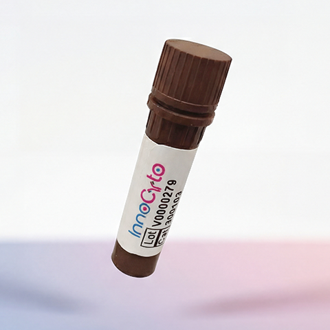
| Cat # | Size | Price | Quantity | |
|---|---|---|---|---|
| 107101 | 100 ug | $80 | ||
| 107102 | 500 ug | $220 |
| Clone | JOVI-3 |
|---|---|
| Application | Flow Cytometry |
| Reactivity | Human |
| Format | Purified |
| Target Name | TCRbV3.1 |
| Isotype | Mouse IgG2a |
| Antibody Type | Monoclonal |
| Regulatory Status | RUO |
| Formulation | Phosphate-buffered solution, pH 7.2, containing 0.09% sodium azide |
| Protein Concentration | 0.5 mg/mL |
| Storage and Handling | The antibody solution should be stored between 2°C and 8°C |
| Recommended Usage | For flow cytometric staining, it is recommended to use less than 0.2 µg of this reagent per 0.5-1.0 million cells in a 100 µL volume. Optimal reagent performance should be determined by titration for each specific application. |
| RRID | AB_3738757 |
| Research Areas | T cells, T cell Activation, Adaptive Immunity |
| See All Formats | Clone JOVI-3 |
TCRβ V3.1, more formally designated TRBV3-1, is a variable gene segment of the T cell receptor (TCR) β chain that contributes to antigen recognition by αβ T lymphocytes. The TCR is central to adaptive immunity, enabling T cells to recognize peptide antigens presented by major histocompatibility complex (MHC) molecules. Use of the TRBV3-1 gene segment defines a subset of T cells with shared structural features in the variable region of their TCR β chain, contributing to repertoire diversity and antigen specificity.
Structurally, TCRβ V3.1 encodes part of the extracellular variable domain of the TCR β chain. During T cell development in the thymus, the TRBV3-1 gene recombines with diversity (Dβ) and joining (Jβ) gene segments through V(D)J recombination. This process, along with junctional diversity, generates a highly variable complementarity-determining region 3 (CDR3), which is the primary determinant of antigen specificity. The resulting TCR β chain pairs with a TCR α chain to form the complete αβ TCR, which associates with the CD3 signaling complex to transduce activation signals. The functional “ligands” of TCRβ V3.1-containing TCRs are peptide-MHC complexes displayed on antigen-presenting cells. Antigen recognition is mediated through interactions between the TCR variable domains and both the peptide and the MHC molecule. In addition to conventional peptide antigens, certain TCR Vβ families, including TRBV3-1, can be selectively engaged by bacterial or viral superantigens. Superantigens bind outside the conventional peptide-binding groove, cross-linking specific TCR Vβ regions with MHC class II molecules and triggering massive, non-specific T cell activation.
TCRβ V3.1 has been implicated in disease primarily through skewed or clonal expansion of TRBV3-1-expressing T cells. Such expansions have been reported in settings of superantigen exposure, chronic infection, autoimmune disease, and some T cell leukemias or lymphomas, where restricted TCR Vβ usage can reflect antigen-driven or malignant proliferation. Monitoring TRBV3-1 usage is therefore useful in studying immune dysregulation and T cell clonality.
In therapeutic and research contexts, TCRβ V3.1 is mainly used as a biomarker rather than a direct drug target. Antibodies specific for TCR Vβ families enable detailed immune repertoire analysis by flow cytometry, aiding in the diagnosis of T cell malignancies and the study of antigen-specific immune responses. In adoptive T cell therapies and TCR-engineered approaches, understanding Vβ usage, including TRBV3-1, contributes to safety assessment and optimization of TCR specificity and function.
Mouse IgG2a Isotype Control Antibody
Anti-Human TCRbV3.1(TRbV28) Antibody TDS
iF488 Anti-Human TCR Vb13.1 Antibody, Clone H131
Have a product or application question? Consult our FAQs or contact us.