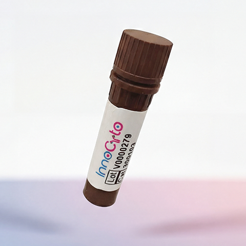
| Cat # | Size | Price | Quantity | |
|---|---|---|---|---|
| 208501 | 25 µg | $45 | ||
| 208502 | 100 µg | $90 |
| Clone | m183AH1 |
|---|---|
| Application | Flow Cytometry |
| Reactivity | Mouse |
| Format | Purified |
| Target Name | CD183, CXCR3, G protein-coupled receptor 9 (GPR9), IP10 receptor (IP10-R), Mig receptor (Mig-R ), CKR-L2 |
| Isotype | Human IgG1 |
| Antibody Type | Monoclonal |
| Regulatory Status | RUO |
| Formulation | Phosphate-buffered solution, pH 7.2, containing 0.09% sodium azide |
| Protein Concentration | 0.5 mg/mL |
| Storage & Handling | The antibody solution should be stored between 2°C and 8°C |
| Recommended Usage | For flow cytometric staining, it is recommended to use less than 0.2 µg of this reagent per 0.5-1.0 million cells in a 100 µL volume. Optimal reagent performance should be determined by titration for each specific application |
| See All Formats | Clone m183AH1 |
CD183, also known as CXCR3, is a chemokine receptor that plays an important role in immune-cell trafficking and inflammatory responses. It is expressed primarily on activated T lymphocytes, particularly Th1 cells and cytotoxic T cells, as well as on natural killer (NK) cells and certain other immune-cell populations. By sensing chemokines produced at sites of inflammation, CXCR3 guides immune cells toward infected tissues, inflamed organs, and tumors, helping coordinate cellular immune responses.
CXCR3 is a seven-transmembrane G protein-coupled receptor (GPCR) belonging to the chemokine receptor family. Its extracellular regions recognize chemokine ligands, while its intracellular domains interact with signaling proteins to regulate cell migration, adhesion, and activation. The principal ligands of CXCR3 are CXCL9 (MIG), CXCL10 (IP-10), and CXCL11 (I-TAC). These chemokines are commonly induced by interferon signaling and bind CXCR3 to activate pathways that promote chemotaxis and other immune functions. Ligand binding can also influence receptor internalization and signaling dynamics.
CXCR3 contributes to host defense against viral and intracellular bacterial infections by recruiting effector lymphocytes to affected tissues. However, excessive or persistent CXCR3 signaling may contribute to inflammatory and autoimmune diseases, including type 1 diabetes, multiple sclerosis, rheumatoid arthritis, and certain forms of tissue inflammation. In cancer, CXCR3 has context-dependent effects: it can promote the recruitment of tumor-killing T cells, but CXCR3 signaling in tumor cells or other microenvironmental cells may also support migration or metastasis in some cancers.
Therapeutically, CXCR3 is being investigated as a target for modulating immune-cell recruitment. Blocking CXCR3 or its chemokine ligands may help reduce damaging inflammation and autoimmune activity. Conversely, enhancing CXCR3-mediated trafficking or engineering immune cells with optimized CXCR3 expression may improve the localization of antitumor T cells within tumors. Its therapeutic value therefore depends on the disease context, the expressing cell type, and the balance between protective immunity and pathological inflammation.
Human IgG1 Isotype Control Antibody
Anti-mouse CD183 (CXCR3) Antibody TDS
Have a product or application question? Consult our FAQs or contact us.