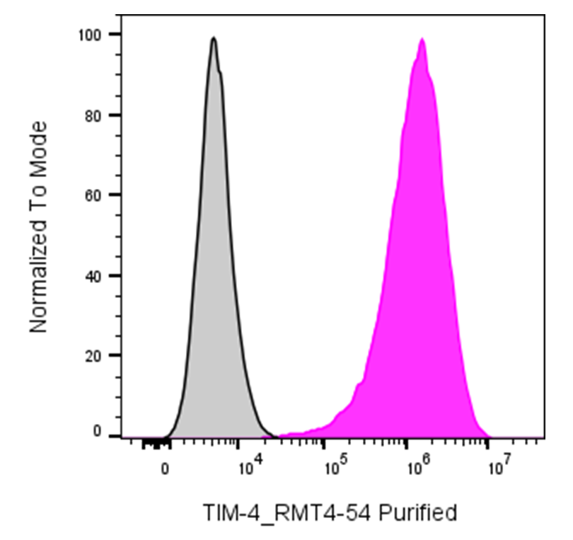
| Cat # | Size | Price | Quantity | |
|---|---|---|---|---|
| 208301 | 25 µg | $65 | ||
| 208302 | 100 µg | $135 |
| Clone | RMT4-54 |
|---|---|
| Application | Flow Cytometry |
| Reactivity | Mouse |
| Format | Purified |
| Target Name | Tim-4, Tim4, T cell immunoglobulin and mucin domain containing protein-4 |
| Isotype | Rat IgG2a |
| Antibody Type | Monoclonal |
| Regulatory Status | RUO |
| Formulation | Phosphate-buffered solution, pH 7.2, containing 0.09% sodium azide |
| Protein Concentration | 0.5 mg/mL |
| Storage & Handling | The antibody solution should be stored between 2°C and 8°C |
| Recommended Usage | For flow cytometric staining, it is recommended to use less than 0.2 µg of this reagent per 0.5-1.0 million cells in a 100 µL volume. Optimal reagent performance should be determined by titration for each specific application |
| See All Formats | Clone RMT4-54 |
Mouse TIM-4 (T-cell immunoglobulin and mucin-domain containing protein 4), encoded by the Havcr2 gene, is a cell-surface immunoregulatory protein expressed primarily on antigen-presenting cells, including macrophages and dendritic cells. TIM-4 plays an important role in the recognition and clearance of apoptotic cells and in the regulation of immune responses. By binding phosphatidylserine exposed on the surface of apoptotic cells, TIM-4 helps phagocytic cells identify and remove dying cells, thereby contributing to tissue homeostasis and limiting the persistence of cellular debris and inflammatory signals.
Mouse TIM-4 is a type I transmembrane glycoprotein belonging to the TIM family. Its extracellular region contains an immunoglobulin-like V-type domain followed by a mucin-like domain, a transmembrane region, and a cytoplasmic tail. Unlike several other TIM family members, TIM-4 lacks a conventional intracellular signaling motif and primarily functions as a ligand-binding and cell-interaction molecule. Its best-characterized ligand is phosphatidylserine (PtdSer), which becomes exposed on the outer leaflet of the plasma membrane during apoptosis. TIM-4 can also participate in interactions with other immune-cell molecules, including pathways involving TIM-1.
Abnormal TIM-4-mediated recognition of apoptotic cells has been implicated in autoimmune and inflammatory diseases. Efficient clearance of apoptotic cells is important for preventing the accumulation of cellular material that can stimulate inappropriate immune responses. TIM-4 expression and function have been investigated in experimental models of autoimmune disease, allergy, transplantation, and tumor immunity. Depending on the cellular and disease context, TIM-4-mediated interactions can either promote immune tolerance or influence inflammatory responses.
Because TIM-4 participates in apoptotic-cell clearance and immune regulation, it is being investigated as a potential therapeutic target. Modulating TIM-4 activity may influence macrophage-mediated clearance, immune tolerance, and inflammatory responses. Experimental approaches targeting the TIM-4 pathway have been explored in autoimmune disease, transplantation, and cancer research. Mouse TIM-4 is therefore also an important research tool for studying phagocytosis, immune regulation, and mechanisms controlling the response to apoptotic cells.
Rat IgG2a Isotype Control Antibody
Have a product or application question? Consult our FAQs or contact us.