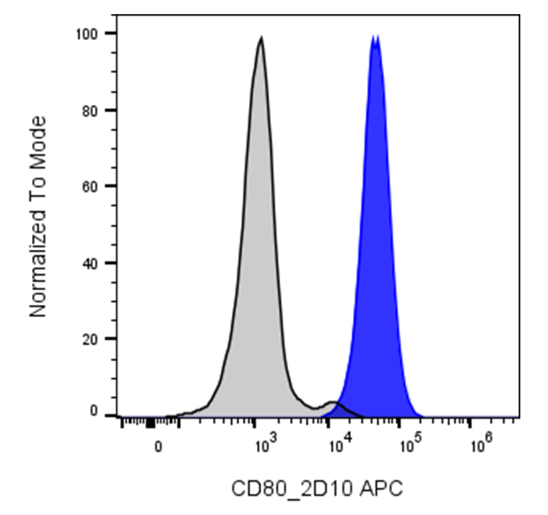
| Cat # | Size | Price | Quantity | |
|---|---|---|---|---|
| 113709 | 25 tests | $90 | ||
| 113710 | 100 tests | $215 |
| Clone | 2D10 |
|---|---|
| Application | Flow Cytometry |
| Reactivity | Human |
| Format | APC |
| Target Name | CD80, B1, B7-1, BB1 |
| Isotype | Mouse IgG1 |
| Antibody Type | Monoclonal |
| Regulatory Status | RUO |
| Formulation | Phosphate-buffered solution, pH 7.2, containing 0.09% sodium azide and 0.2% (w/v) BSA |
| Protein Concentration | Supplied at a lot-specific concentration. |
| Storage & Handling | The antibody solution should be stored undiluted between 2°C and 8°C, and protected from prolonged exposure to light. Do not freeze. |
| Recommended Usage | For flow cytometric staining, it is recommended to use 5 µL of this reagent per 0.5-1.0 million cells in a 100 µL volume. Optimal reagent performance should be determined by titration for each specific application. FITC has an excitation max at 493 nm and an emission max at 525 nm. |
| Excitation Laser | Red Laser (633 nm) |
| See All Formats | Clone 2D10 |
Human :contentReference[oaicite:0]{index=0}, also known as B7-1, is a key co-stimulatory molecule expressed primarily on professional antigen-presenting cells such as dendritic cells, B cells, and macrophages. It plays a central role in regulating T cell activation by providing the “second signal” required for full immune activation, complementing T cell receptor (TCR) recognition of antigen.
Structurally, CD80 is a type I transmembrane glycoprotein and a member of the immunoglobulin superfamily. It consists of two extracellular immunoglobulin-like domains (one V-like and one C-like), a transmembrane region, and a short cytoplasmic tail. CD80 can form homodimers on the cell surface, which may enhance its binding avidity and functional activity during immune synapse formation.
Functionally, CD80 interacts with two primary ligands on T cells: :contentReference[oaicite:1]{index=1} and :contentReference[oaicite:2]{index=2}. Binding to CD28 delivers a co-stimulatory signal that promotes T cell proliferation, cytokine production, and survival. In contrast, engagement with CTLA-4 transmits inhibitory signals that suppress T cell responses and maintain immune homeostasis. Thus, CD80 is a critical regulator balancing immune activation and inhibition.
In disease, dysregulation of CD80 expression or signaling is implicated in autoimmunity, chronic infections, and cancer. Overactivation of CD80-CD28 signaling can contribute to autoimmune disorders, while tumors may exploit the CD80–CTLA-4 pathway to suppress anti-tumor immunity.
Therapeutically, CD80 is indirectly targeted in immunotherapy. Agents such as :contentReference[oaicite:3]{index=3} block CTLA-4, enhancing CD80/CD28-mediated T cell activation. Additionally, engineered cells expressing CD80 are being explored to boost immune responses in cancer and vaccine strategies.
APC Mouse IgG1 Isotype Control Antibody
APC Anti-Human CD80 Antibody TDS
Have a product or application question? Consult our FAQs or contact us.