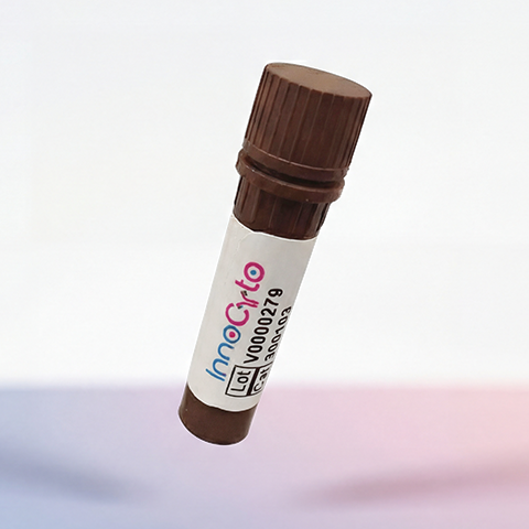
| Cat # | Size | Price | Quantity | |
|---|---|---|---|---|
| 809703 | 25 ug | $245 | ||
| 809704 | 100 ug | $595 |
| Application | Flow Cytometry |
|---|---|
| Format | Liquid, APC |
| Expression Host | CHO |
| Target Name | CD33, SIGLEC3, gp67 |
| Species | Human |
| Sources | Recombinant Human CD33 Protein (Asp18-His259) with C-terminus Fc-tag is expressed in CHO cell and conjugated to APC. |
| Accession Number | P20138 |
| Molecular Weight | The protein has a predicted molecular weight of 53 kDa. Under DTT-reducing conditions, it migrates at approximately 70-90 kDa on SDS-PAGE prior to conjugation. |
| Affinity Tag | C-Fc |
| Regulatory Status | RUO |
| Formulation | 1xPBS buffer, pH7.4, 0.09% NaN3 with a carrier protein |
| Endotoxin level | Not tested |
| Protein Concentration | 25µg size is bottled at 0.1mg/mL concentration. 100 µg size is bottled at lot specific concentration. |
| Storage and Handling | Briefly centrifuge the vial upon receipt. An unopened vial may be stored at 2–8°C for up to six months. |
| Research Areas | Monocytes, Macrophages, Dendritic cells, Innate Immunity, Cancer Marker |
CD33, also known as Siglec-3 (sialic acid-binding immunoglobulin-like lectin 3), is a transmembrane receptor belonging to the Siglec family of proteins that plays an important role in regulating immune cell function. CD33 is predominantly expressed on myeloid lineage cells, including monocytes, macrophages, granulocytes, and myeloid dendritic cells, as well as on myeloid progenitor cells in the bone marrow. The protein functions as an inhibitory receptor that modulates immune responses by dampening cellular activation signals. Upon ligand binding, CD33 recruits phosphatases that suppress inflammatory signaling pathways, thereby maintaining immune homeostasis and preventing excessive immune activation.
Structurally, CD33 is a type I transmembrane glycoprotein of approximately 67 kDa consisting of an extracellular region with two immunoglobulin-like domains (one V-set and one C2-set domain), a single transmembrane domain, and a cytoplasmic tail containing immunoreceptor tyrosine-based inhibitory motifs (ITIMs). The V-set domain at the N-terminus contains the sialic acid-binding site responsible for ligand recognition. The cytoplasmic ITIMs, when phosphorylated, recruit SHP-1 and SHP-2 phosphatases that deliver inhibitory signals to suppress cell activation, proliferation, and cytokine production. This structural organization enables CD33 to function as a checkpoint receptor that fine-tunes myeloid cell responses.
CD33 binds to sialylated glycans, particularly α2,6-linked and α2,3-linked sialic acids, which are present on glycoproteins and glycolipids on cell surfaces and in the extracellular matrix. These sialic acid-containing ligands are widely distributed throughout the body, allowing CD33 to recognize "self" markers and maintain immune tolerance. The protein's ability to recognize sialylated structures makes it an important regulator of innate immunity and inflammation.
In disease contexts, CD33 is highly significant in acute myeloid leukemia (AML), where it is expressed on leukemic blasts in approximately 85-90% of cases. This expression pattern has made CD33 an attractive therapeutic target. Gemtuzumab ozogamicin, an antibody-drug conjugate targeting CD33, was the first CD33-directed therapy approved for AML treatment, delivering a cytotoxic payload specifically to CD33-positive leukemic cells. Additionally, CD33 genetic variants have been associated with Alzheimer's disease risk, as the protein is expressed on microglia and may influence neuroinflammation and amyloid-beta clearance. Beyond gemtuzumab ozogamicin, newer CD33-targeted therapies under development include bispecific antibodies, CAR-T cells, and next-generation antibody-drug conjugates, establishing CD33 as a key target in hematologic malignancy treatment and potentially in neurodegenerative disease modulation.
APC Human CD33 Protein (C-Fc) TDS
Biotin Anti-Human CD14 Antibody, Clone 63D3
PE Anti-Human CD14 Antibody, Clone 63D3
Anti-Human CD14 Antibody, Clone M5E2
iF647 Anti-Human CD14 Antibody, Clone M5E2
Anti-Human CD33 (Gemtuzumab Biosimilar), Clone Gemtuzumab
Human CD33 Protein (C-His-Avi)
Biotin Human CD33 Protein (C-His-Avi)
Have a product or application question? Consult our FAQs or contact us.