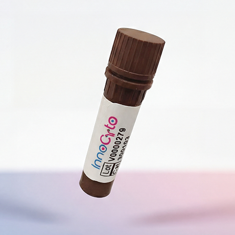
| Cat # | Size | Price | Quantity | |
|---|---|---|---|---|
| 802703 | 25 ug | $245 | ||
| 802704 | 100 ug | $595 |
| Application | Flow Cytometry |
|---|---|
| Format | Liquid, APC |
| Expression Host | CHO |
| Target Name | FOLR-1, FBP, FOLR, FRα |
| Species | Human |
| Sources | Human FOLR1 (Arg25-Met233) with C-terminus His Tag is expressed in CHO cells and conjugated to APC. |
| Accession Number | P15328 |
| Molecular Weight | The protein has a predicted molecular weight of 28.6 kDa. Under DTT-reducing conditions, it migrates at approximately 35-45 kDa on SDS-PAGE prior to conjugation. |
| Affinity Tag | C-His |
| Regulatory Status | RUO |
| Formulation | 1xPBS buffer, pH7.4, 0.09% NaN3 with a carrier protein |
| Endotoxin level | Not tested |
| Protein Concentration | 25µg size is bottled at 0.1mg/mL concentration. 100 µg size is bottled at lot specific concentration. |
| Storage and Handling | Briefly centrifuge the vial upon receipt. An unopened vial may be stored at 2–8°C for up to six months. |
| Research Areas | Epithelial cells, Cancer Marker |
Folate receptor alpha (FOLR1), also known as FRα or folate receptor 1, is a high-affinity, glycosylphosphatidylinositol (GPI)-anchored cell surface protein primarily responsible for binding and internalizing folate and its derivatives. Folates are essential vitamins that participate in one-carbon metabolism, supporting nucleotide synthesis and methylation reactions crucial for DNA replication and repair. FOLR1 mediates cellular uptake of folate through receptor-mediated endocytosis, complementing the activity of other folate transporters such as the reduced folate carrier (RFC) and the proton-coupled folate transporter (PCFT).
Structurally, FOLR1 is a glycoprotein of approximately 38–42 kDa, composed of a single extracellular domain anchored to the plasma membrane via a GPI linkage. The extracellular domain contains a hydrophobic binding pocket that specifically recognizes and binds reduced folate and folate analogs with nanomolar affinity. Structural studies have revealed that this pocket is formed by β-sheets and loop regions that precisely accommodate the pterin and glutamate moieties of folate. The protein’s GPI anchor localizes it to lipid rafts, membrane microdomains involved in signaling and endocytosis, thereby facilitating efficient internalization and trafficking of folate-loaded vesicles.
FOLR1 has a particularly high expression in certain normal tissues, including the kidneys, placenta, and choroid plexus, but its expression in most normal epithelial tissues is limited. In contrast, FOLR1 is markedly overexpressed in several epithelial-derived cancers, such as ovarian, endometrial, breast, non–small cell lung, and renal carcinomas. This differential expression pattern plays a role in oncogenesis, as tumor cells exploit high folate uptake to support their increased proliferative demands. Moreover, FOLR1 expression correlates with tumor aggressiveness and poor prognosis in some cancers, making it a valuable biomarker for diagnosis and targeted therapy.
Therapeutically, FOLR1 serves as an attractive target for cancer treatment due to its selective tumor overexpression. Strategies exploiting this receptor include folate–drug conjugates (e.g., vintafolide), antibody–drug conjugates (ADCs) such as mirvetuximab soravtansine, and folate-based imaging agents for tumor detection. These therapies deliver cytotoxic agents specifically to FOLR1-positive cancer cells, minimizing off-target effects. Clinical trials have shown promising efficacy, particularly in ovarian cancers with high FOLR1 expression, establishing the receptor as a key biomarker and therapeutic target in precision oncology.
APC Human FOLR1 Protein (C-His) TDS
Human FOLR1 Protein (C-His-Avi)
Biotin Human FOLR1 Protein (C-His-Avi)
PE Human FOLR1 Protein (C-His)
Have a product or application question? Consult our FAQs or contact us.