| Cat # | Size | Price | Quantity | |
|---|---|---|---|---|
| 803303 | 25 ug | $245 | ||
| 803304 | 100 ug | $595 |
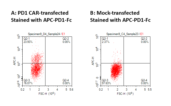
| Application | Flow Cytometry |
|---|---|
| Format | Liquid, APC |
| Expression Host | HEK293 |
| Target Name | PD1, PDCD1, CD279, SLEB2 |
| Species | Human |
| Sources | Human PD-1 protein (NP_005009.2) (Leu25-Gln167) with C-terminus Fc tag is expressed in HEK293 cells and conjugated to APC |
| Accession Number | Q15116 |
| Molecular Weight | The protein has a predicted molecular weight of 19 kDa. Under DTT-reducing conditions, it migrates at approximately 25-30 kDa on SDS-PAGE prior to conjugation. |
| Affinity Tag | C-Fc |
| Regulatory Status | RUO |
| Formulation | 1xPBS buffer, pH7.4, 0.09% NaN3 with a carrier protein |
| Endotoxin level | Not tested |
| Protein Concentration | 25µg size is bottled at 0.1mg/mL concentration. 100 µg size is bottled at lot specific concentration. |
| Storage and Handling | Briefly centrifuge the vial upon receipt. An unopened vial may be stored at 2–8°C for up to six months. |
| Research Areas | Immune Checkpoints, Activated T cells, Cancer Immunology, Tregs, Tfh cells, Activated B cells |
CD279, also known as Programmed Cell Death Protein 1 (PD-1), is a crucial immune checkpoint receptor that regulates T cell activation and prevents autoimmunity. This transmembrane protein plays a pivotal role in maintaining immune homeostasis by delivering inhibitory signals that dampen excessive immune responses.
PD-1 is a type I transmembrane glycoprotein belonging to the immunoglobulin superfamily. It contains an extracellular immunoglobulin variable (IgV)-like domain, a transmembrane region, and an intracellular tail with two tyrosine-based signaling motifs: an immunoreceptor tyrosine-based inhibitory motif (ITIM) and an immunoreceptor tyrosine-based switch motif (ITSM). When engaged, these motifs recruit phosphatases that inhibit T-cell receptor signaling, effectively suppressing T-cell activation, proliferation, and cytokine production.
PD-1 interacts with two primary ligands: PD-L1 (B7-H1/CD274) and PD-L2 (B7-DC/CD273). PD-L1 is widely expressed on various cell types, including tumor cells, antigen-presenting cells, and non-hematopoietic tissues, while PD-L2 expression is more restricted to antigen-presenting cells. These ligand-receptor interactions serve as critical brakes on immune responses. In cancer, tumor cells exploit the PD-1/PD-L1 pathway to evade immune surveillance. By upregulating PD-L1 expression, tumors effectively "turn off" infiltrating T-cells, preventing effective anti-tumor immunity. This mechanism contributes to tumor progression and immune escape across multiple cancer types.
The discovery of PD-1's role in cancer has revolutionized oncology through immune checkpoint inhibitors. Monoclonal antibodies targeting PD-1 (pembrolizumab, nivolumab) or PD-L1 (atezolizumab, durvalumab) block this inhibitory pathway, reinvigorating anti-tumor T-cell responses. These therapies have demonstrated remarkable success in treating melanoma, non-small cell lung cancer, renal cell carcinoma, and numerous other malignancies, fundamentally transforming cancer treatment paradigms and offering durable responses in previously untreatable cancers.
APC Human PD1 (CD279) Protein (C-Fc) TDS
Anti-Mouse CD274 (PD-L1) Antibody, Clone 10F.9G2
Biotin Anti-Mouse CD274 (PD-L1) Antibody, Clone 10F.9G2
Anti-Human PD-L1 (Atezolizumab Biosimilar), Clone Atezolizuma
Anti-Human PD-L1 (Avelumab Biosimilar), Clone Avelumab
Anti-Human PD1 (Camrelizumab Biosimilar), Clone Camrelizumab
Anti-Human PD1 (Cemiplimab Biosimilar), Clone Cemiplimab
Anti-Human PD1 (Nivolumab Biosimilar), Clone Nivolumab
Anti-Human PD1 (Pembrolizumab Biosimilar), Clone Pembrolizumab
Anti-Human PD1 (Spartalizumab Biosimilar), Clone Spartalizumab
Human PD1 (CD279) Protein (C-His-Avi)
Have a product or application question? Consult our FAQs or contact us.