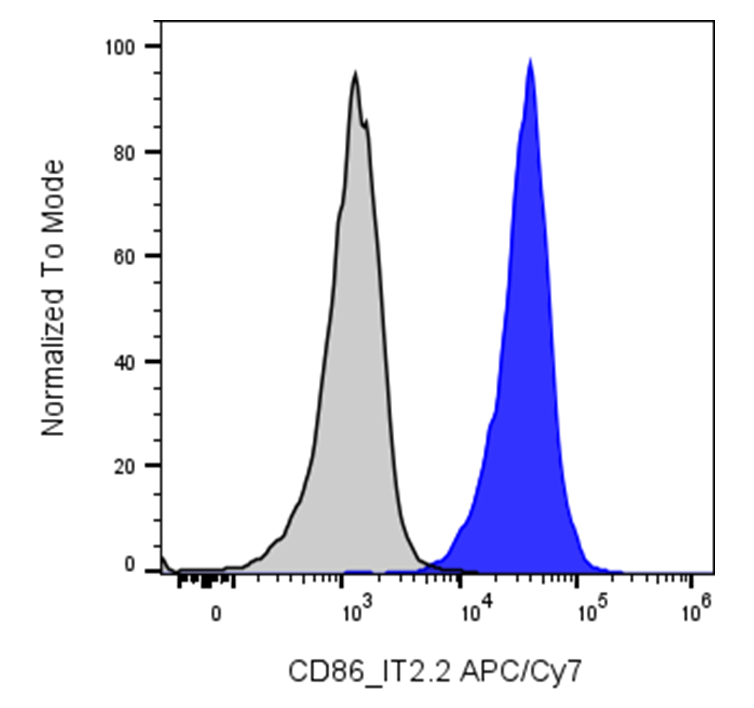
| Cat # | Size | Price | Quantity | |
|---|---|---|---|---|
| 113911 | 25 tests | $140 | ||
| 113912 | 100 tests | $280 |
| Clone | IT2.2 |
|---|---|
| Application | Flow Cytometry |
| Reactivity | Human |
| Format | APC/Cyanine7 |
| Target Name | CD86, B7-2, B70 |
| Isotype | Mouse IgG2b |
| Antibody Type | Monoclonal |
| Regulatory Status | RUO |
| Formulation | Phosphate-buffered solution, pH 7.2, containing 0.09% sodium azide and 0.2% (w/v) BSA |
| Protein Concentration | Supplied at a lot-specific concentration. |
| Storage & Handling | The antibody solution should be stored undiluted between 2°C and 8°C, and protected from prolonged exposure to light. Do not freeze. |
| Recommended Usage | For flow cytometric staining, it is recommended to use 5 µL of this reagent per 0.5-1.0 million cells in a 100 µL volume. Optimal reagent performance should be determined by titration for each specific application. APC/Cyanine7 has an excitation max at 650 nm and an emission max at 774 nm. |
| Excitation Laser | Red Laser (633 nm) |
| See All Formats | Clone IT2.2 |
Human CD86, also known as B7-2, is a type I transmembrane glycoprotein and a key costimulatory molecule expressed primarily on professional antigen-presenting cells (APCs), including dendritic cells, macrophages, and B cells. Encoded by the CD86 gene, CD86 is rapidly upregulated following immune activation and plays an essential role in regulating adaptive immune responses. Its primary function is to provide the second signal required for full T-cell activation, proliferation, and cytokine production after antigen recognition by the T-cell receptor.
CD86 belongs to the immunoglobulin (Ig) superfamily and consists of two extracellular Ig-like domains, a single transmembrane domain, and a short cytoplasmic tail. The principal ligands for CD86 are CD28 and cytotoxic T-lymphocyte-associated protein 4 (CTLA-4), both expressed on T cells. Binding of CD86 to CD28 delivers a positive costimulatory signal that promotes T-cell activation and survival, whereas interaction with CTLA-4 transmits an inhibitory signal that suppresses immune responses and helps maintain peripheral tolerance. The balance between these receptor interactions is critical for immune homeostasis.
Aberrant CD86 expression has been implicated in numerous diseases, including autoimmune disorders such as rheumatoid arthritis, systemic lupus erythematosus, and multiple sclerosis, where excessive costimulation contributes to pathogenic T-cell activation. Increased CD86 expression has also been observed in chronic inflammatory conditions and certain hematologic and solid malignancies, where it may influence antitumor immunity or immune evasion.
CD86 is an important therapeutic target for immune modulation. CTLA-4-Ig fusion proteins, including abatacept and belatacept, bind CD86 (and CD80) to block CD28-mediated costimulation, thereby suppressing T-cell activation. These agents are approved for the treatment of autoimmune diseases and prevention of transplant rejection. Ongoing research is evaluating additional therapeutic strategies targeting the CD80/CD86 pathway to enhance cancer immunotherapy, promote transplantation tolerance, and treat autoimmune and inflammatory diseases.
APC/Cyanine7 Mouse IgG2b Isotype Control
APC/Cyanine7 Anti-Human CD86 Antibody TDS
Have a product or application question? Consult our FAQs or contact us.