| Cat # | Size | Price | Quantity | |
|---|---|---|---|---|
| 200717 | 25 tests | $80 | ||
| 200718 | 100 tests | $240 |
| Clone | 1A8 |
|---|---|
| Application | Flow Cytometry |
| Reactivity | Mouse |
| Format | APC/Cyanine7 |
| Target Name | Ly-6G, Lymphocyte antigen 6 complex, locus G |
| Isotype | Rat IgG2a |
| Antibody Type | Monoclonal |
| Regulatory Status | RUO |
| Formulation | Phosphate-buffered solution, pH 7.2, containing 0.09% sodium azide and 0.2% (w/v) BSA |
| Protein Concentration | Supplied at a lot-specific concentration. |
| Storage&Handling | The antibody solution should be stored undiluted between 2°C and 8°C, and protected from prolonged exposure to light. Do not freeze. |
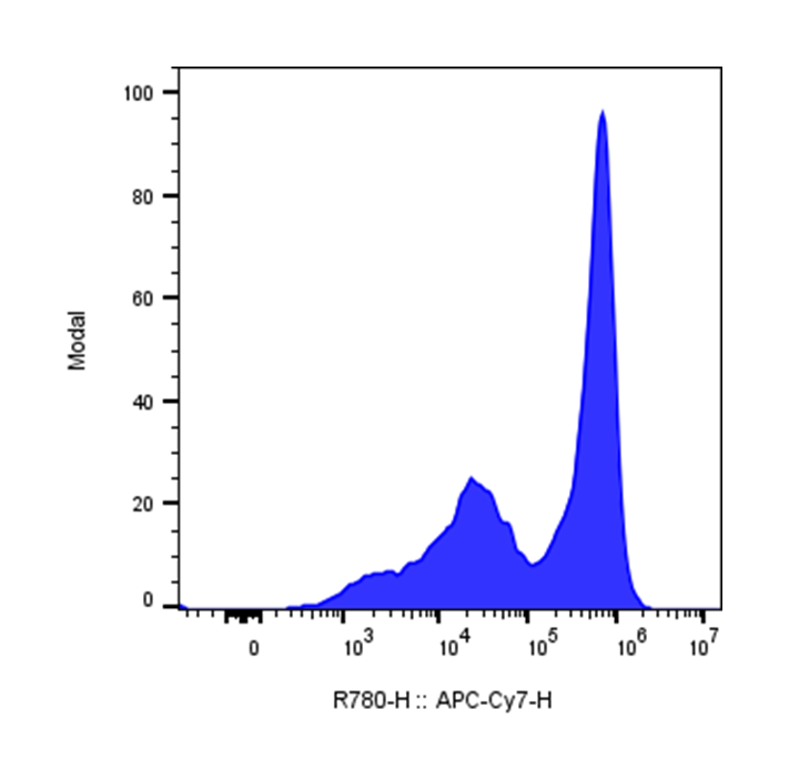
| Recommended Usage | For flow cytometric staining, it is recommended to use 5 µL of this reagent per 0.5-1.0 million cells in a 100 µL volume. Optimal reagent performance should be determined by titration for each specific application. APC/Cyanine7 has an excitation max at 650 nm and an emission max at 774 nm. |
| Excitation Laser | Red Laser (633 nm) |
| Research Areas | Neutrophils, plasmacytoid Dendritic cells |
| See All Formats | Clone 1A8 |
Lymphocyte antigen 6 complex, locus G (Ly-6G), is a 21-25 kDa glycosylphosphatidylinositol (GPI)-anchored surface protein. In mice, it is expressed predominantly on peripheral granulocytes, especially neutrophils, and on the majority of myeloid cells in the bone marrow. Ly-6G is transiently detected on developing monocytes but is stably and highly expressed on mature neutrophils. It plays important roles in neutrophil infiltration, recruitment, and migration, and associates with β2 integrins CD11a and CD11b to regulate their expression and function. The natural ligand for Ly-6G remains unknown, and it is widely used as a specific marker for murine neutrophils in immunological studies.
APC/Cyanine7 Rat IgG2a Isotype Control Antibody
APC/Cyanine7 Anti-Mouse Ly-6G Antibody TDS
iF647 Anti-Mouse CD11a Antibody, Clone M17/4
PE Anti-Mouse/Human CD11b (Integrin αM) Antibody, Clone M1/70
Have a product or application question? Consult our FAQs or contact us.