| Cat # | Size | Price | Quantity | |
|---|---|---|---|---|
| 108903 | 25 ug | $70 | ||
| 108904 | 100 ug | $180 |
| Clone | THB5 |
|---|---|
| Application | Flow Cytometry |
| Reactivity | Human |
| Format | Biotin |
| Target Name | CD21, Complement C3d receptor (C3dR), complement receptor 2 (CR2), Epstein-Barr virus receptor |
| Isotype | Mouse IgG2a |
| Antibody Type | Monoclonal |
| Regulatory Status | RUO |
| Formulation | Phosphate-buffered solution, pH 7.2, containing 0.09% sodium azide and 0.2% (w/v) BSA |
| Protein Concentration | 0.2 mg/mL |
| Storage and Handling | The antibody solution should be stored undiluted between 2°C and 8°C, and protected from prolonged exposure to light. Do not freeze. |
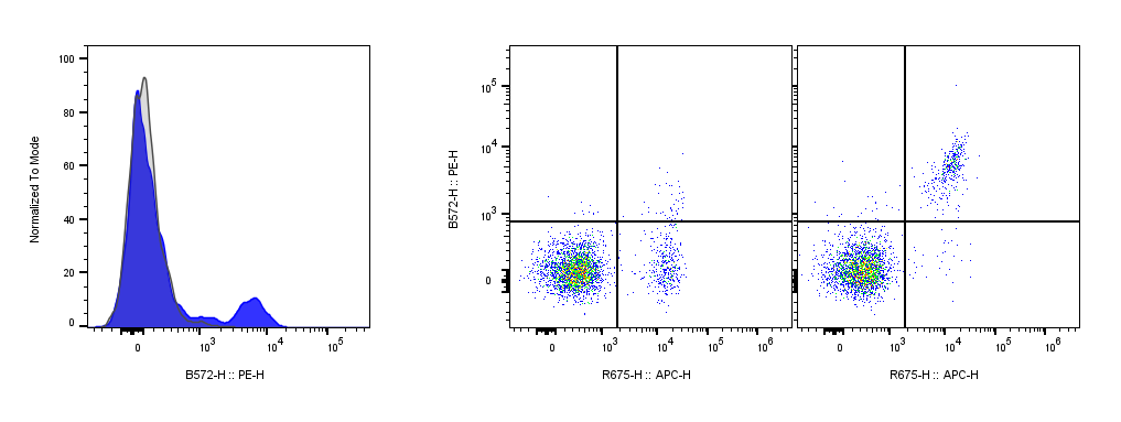
| Recommended Usage | For flow cytometric staining, it is recommended to use less than 0.1 µg of this reagent per 0.5-1.0 million cells in a 100 µL volume. Optimal reagent performance should be determined by titration for each specific application. For detection, use a secondary reagent with this product. |
| RRID | AB_3738867 |
| Research Areas | B cells, follicular Dendritic cells, EBV, Infectious Diseases |
| See All Formats | Clone THB5 |
CD21, also known as complement receptor 2 (CR2), is a surface glycoprotein expressed primarily on mature B cells, follicular dendritic cells, and certain epithelial cells. It serves as a receptor for complement fragments and viral particles, playing a central role in regulating adaptive immune responses. CD21 is essential for the activation and maturation of B cells, helping to lower the threshold for B cell receptor (BCR) signaling by binding complement-tagged antigens, thereby enhancing antibody production and immune memory formation.
Structurally, CD21 is a type I transmembrane protein of approximately 145 kDa and belongs to the regulators of complement activation (RCA) family. It comprises 15 or 16 extracellular short consensus repeats (SCRs), each containing about 60 amino acid residues stabilized by disulfide bonds. These SCR domains contribute to ligand recognition and binding, while the short cytoplasmic tail participates in intracellular signaling through interactions with other surface molecules, such as CD19 and CD81, forming the B cell co-receptor complex. This multimolecular complex plays a critical role in amplifying B cell activation in response to antigen engagement.
The principal ligands for CD21 are complement fragments C3d, iC3b, and C3dg, which are generated during complement activation. CD21 also acts as a receptor for the Epstein-Barr virus (EBV) glycoprotein gp350/220, facilitating viral entry into B cells and establishing latent infection. This dual function - immune modulation and viral susceptibility - underpins its significance in both health and disease.
CD21 involvement in disease extends beyond viral infection. Dysregulation of CD21 expression or signaling contributes to immune-related disorders such as systemic lupus erythematosus (SLE), where altered receptor levels impair clearance of immune complexes and promote autoreactivity. In chronic lymphocytic leukemia (CLL) and some lymphomas, CD21 expression serves as a diagnostic and phenotypic marker for subtyping B cell malignancies. Conversely, its engagement by EBV plays a role in the development of EBV-associated cancers such as Burkitt lymphoma and nasopharyngeal carcinoma.
Therapeutically, CD21 offers potential as a target for modulating immune function and managing infection or autoimmunity. Inhibition of CD21-Epstein-Barr virus interactions represents a strategy for preventing viral infection, while modulation of the CD21-C3d pathway may fine-tune B cell responses in autoimmune and vaccine contexts. Ongoing studies continue to define how manipulating CD21 signaling could optimize immunotherapy and immune tolerance.
Biotin Mouse IgG2a Isotype Control Antibody
Biotin Anti-Human CD21 Antibody TDS
Anti-Human CD19 Antibody, Clone HIB19
iF560 Anti-Human CD19 Antibody, Clone HIB19
iF647 Anti-Human CD19 Antibody, Clone 019AM2b
Biotin Anti-Human CD19 Antibody, Clone 019AM2b
iF405 Anti-Human CD81 Antibody, Clone 5A6
PE Anti-Human CD81 Antibody, Clone 5A6
Have a product or application question? Consult our FAQs or contact us.