| Cat # | Size | Price | Quantity | |
|---|---|---|---|---|
| 814003 | 25 ug | $245 | ||
| 814004 | 100 ug | $595 |
| Application | ELISA, BLI |
|---|---|
| Format | Liquid, Biotinylated |
| Expression Host | CHO |
| Target Name | B7-H5, SISP1, Gi24, VISTA |
| Species | Human |
| Sources | Recombinant Human Vista (Phe33-Ala194) with C-terminus Fc-Avi-tag is expressed in CHO cell. This protein was site-specifically labeled with Biotin by BirA ligase. |
| Accession Number | Q9H7M9 |
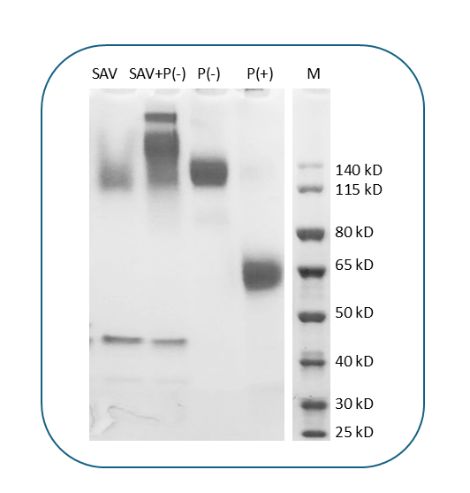
| Molecular Weight | The protein has a predicted molecular weight of 47 kDa. Under DTT-reducing conditions, it migrates at approximately 55-65 kDa on SDS-PAGE. |
| Affinity Tag | C-Fc-Avi |
| Purity | >95% based on SDS-PAGE under reducing condition |
| Regulatory Status | RUO |
| Formulation | 1xPBS buffer, pH7.4, 0.22 µm filtered |
| Endotoxin level | Not tested |
| Protein Concentration | 25µg size is bottled at 0.2mg/mL concentration. 100 µg size is supplied at a lot-specific concentration. |
| Storage and Handling | Briefly centrifuge the vial upon receipt. An unopened vial can be stored at 4°C for up to 2 weeks, or at -20°C or below for up to six months. The protein may be further diluted to 0.1 mg/mL using 0.22 µm-filtered PBS buffer (pH 7.4). For long-term storage, the diluted stock solution should be aliquoted and stored at ≤ –70°C to minimize freeze-thaw cycles. If additional dilution is required, carrier proteins such as FBS or BSA should be added to maintain protein stability. |
| Recommended Usage | For detection, use a secondary reagent with this product. |
VISTA (V-domain Ig suppressor of T cell activation), also known as PD-1H or B7-H5, is an immune checkpoint protein that functions as a negative regulator of T cell activation and immune responses. VISTA is predominantly expressed on hematopoietic cells, particularly myeloid cells such as macrophages, dendritic cells, and neutrophils, as well as on CD4+ and CD8+ T cells and regulatory T cells. The protein plays a crucial role in maintaining immune homeostasis by suppressing T cell proliferation, cytokine production, and effector functions. VISTA exhibits unique pH-dependent binding properties, with enhanced activity in acidic environments such as the tumor microenvironment and inflamed tissues, making it particularly effective at suppressing immune responses in these contexts.
Structurally, VISTA is a type I transmembrane protein of approximately 30-35 kDa belonging to the immunoglobulin superfamily. The extracellular region contains a single IgV-like domain that mediates ligand interactions, distinguishing it from other B7 family members that typically have both IgV and IgC domains. The protein features a unique arrangement of histidine residues in its extracellular domain that confer pH sensitivity, allowing VISTA to function optimally at acidic pH (around 6.0-6.5) found in tumor and inflammatory microenvironments. VISTA also contains a transmembrane domain and a cytoplasmic tail that may participate in intracellular signaling, though its signaling mechanisms are still being elucidated. The protein can function both as a ligand and as a receptor, providing bidirectional immune regulation.
The ligand-receptor relationships for VISTA are complex and still being investigated. VISTA has been reported to interact with multiple binding partners, including VSIG-3 (V-set and immunoglobulin domain-containing 3), also known as IGSF11, which serves as a receptor for VISTA on T cells. Additionally, VISTA may engage in homophilic interactions (VISTA-VISTA binding) and has been suggested to interact with PSGL-1 (P-selectin glycoprotein ligand-1) under certain conditions. The pH-dependent nature of these interactions adds complexity to VISTA's regulatory functions, with binding affinity modulated by the local microenvironment.
In disease contexts, VISTA plays a significant role in cancer immune evasion and autoimmunity. VISTA is upregulated in various tumors, including melanoma, ovarian cancer, pancreatic cancer, and colorectal cancer, where it contributes to creating an immunosuppressive tumor microenvironment that inhibits antitumor T cell responses. High VISTA expression correlates with poor prognosis and resistance to other immune checkpoint therapies in several cancer types. Conversely, VISTA deficiency or blockade has been associated with enhanced susceptibility to autoimmune diseases, highlighting its role in maintaining immune tolerance. Therapeutically, VISTA is emerging as a promising target for cancer immunotherapy. Multiple anti-VISTA antibodies are in clinical development, including CI-8993 and HMBD-002, which aim to block VISTA's inhibitory function and restore antitumor immunity. These agents are being tested both as monotherapies and in combination with other checkpoint inhibitors such as anti-PD-1/PD-L1 antibodies. Additionally, VISTA agonists are being explored for treating autoimmune and inflammatory diseases by enhancing immune suppression. The unique pH-dependent activity of VISTA makes it an attractive target for selectively modulating immunity in acidic disease microenvironments while minimizing systemic effects.
Biotin Human Vista Protein (C-Fc-Avi) TDS
Human Vista Protein (C-Fc-Avi)
APC Human Vista Protein (C-Fc)
Have a product or application question? Consult our FAQs or contact us.