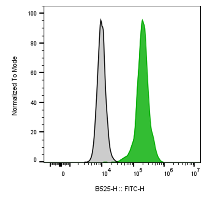
| Cat # | Size | Price | Quantity | |
|---|---|---|---|---|
| 117809 | 25 tests | $75 | ||
| 117810 | 100 tests | $150 |
| Clone | 011cAM1 |
|---|---|
| Application | Flow Cytometry |
| Reactivity | Human |
| Format | FITC |
| Target Name | CD11c, p150Integrin αx subunit, ITGAX, CR4 |
| Isotype | Mouse IgG1 |
| Antibody Type | Monoclonal |
| Regulatory Status | RUO |
| Formulation | Phosphate-buffered solution, pH 7.2, containing 0.09% sodium azide and 0.2% (w/v) BSA |
| Protein Concentration | Supplied at a lot-specific concentration. |
| Storage & Handling | The antibody solution should be stored undiluted between 2°C and 8°C, and protected from prolonged exposure to light. Do not freeze. |
| Recommended Usage | For flow cytometric staining, it is recommended to use 5 µL of this reagent per 0.5-1.0 million cells in a 100 µL volume. Optimal reagent performance should be determined by titration for each specific application. FITC has an excitation max at 493 nm and an emission max at 525 nm. |
| Excitation Laser | Blue Laser (488 nm) |
| See All Formats | Clone 011cAM1 |
Human CD11c, also known as integrin alpha-X (ITGAX), is a cell-surface adhesion and immune-regulatory molecule predominantly expressed on dendritic cells, monocytes, macrophages, and subsets of B cells and other myeloid cells. CD11c forms a heterodimer with the integrin β2 subunit, CD18 (ITGB2), to create the complement receptor 4 (CR4) complex. Through interactions with extracellular matrix and immune-associated ligands, CD11c contributes to leukocyte adhesion, migration, phagocytosis, antigen presentation, and regulation of innate and adaptive immune responses.
CD11c is a large type I transmembrane glycoprotein containing an extracellular α-chain of approximately 1,163 amino acids, a single transmembrane region, and a relatively short cytoplasmic tail. Its major functional receptor is the CD11c/CD18 heterodimer. Known ligands include iC3b, fibrinogen, and several cell-surface and extracellular matrix molecules. Engagement of CD11c can regulate cytoskeletal organization, cell adhesion, phagocytosis, and intracellular signaling, although its effects depend strongly on the cellular context and activating signals.
CD11c is particularly important in immune-mediated diseases because it marks and participates in the function of activated myeloid and dendritic-cell populations. Altered CD11c-positive cell populations have been associated with autoimmune diseases such as rheumatoid arthritis, systemic lupus erythematosus, inflammatory bowel disease, and multiple sclerosis. CD11c-positive tumor-associated myeloid cells can also influence tumor immunity, potentially contributing either to anti-tumor responses or immunosuppressive tumor microenvironments.
Therapeutically, CD11c represents an attractive target for selectively modulating dendritic cells and other myeloid populations. Antibody-drug conjugates, antibody-mediated depletion strategies, and CD11c-targeted antigen-delivery systems have been investigated for cancer, autoimmunity, and vaccine development. Rather than simply blocking CD11c signaling, targeted delivery through CD11c may provide a way to selectively manipulate antigen-presenting cells and enhance immune responses.
FITC Mouse IgG1 Isotype Control Antibody
FITC Anti-Human CD11c Antibody TDS
Have a product or application question? Consult our FAQs or contact us.