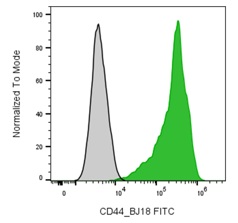
| Cat # | Size | Price | Quantity | |
|---|---|---|---|---|
| 120303 | 25 tests | $75 | ||
| 120304 | 100 tests | $165 |
| Clone | BJ18 |
|---|---|
| Application | Flow Cytometry |
| Reactivity | Human |
| Format | FITC |
| Target Name | CD44, H-CAM, Ly-24, gp85 |
| Isotype | mouse IgG1 |
| Antibody Type | Monoclonal |
| Regulatory Status | RUO |
| Formulation | Phosphate-buffered solution, pH 7.2, containing 0.09% sodium azide and 0.2% (w/v) BSA |
| Protein Concentration | Supplied at a lot-specific concentration. |
| Storage & Handling | The antibody solution should be stored undiluted between 2°C and 8°C, and protected from prolonged exposure to light. Do not freeze. |
| Recommended Usage | For flow cytometric staining, it is recommended to use 5 µL of this reagent per 0.5-1.0 million cells in a 100 µL volume. Optimal reagent performance should be determined by titration for each specific application. FITC has an excitation max at 493 nm and an emission max at 525 nm. |
| Excitation Laser | Blue Laser (488 nm) |
| See All Formats | Clone BJ18 |
Human CD44 is a widely expressed type I transmembrane glycoprotein that functions as a cell adhesion molecule involved in cell-cell and cell-extracellular matrix interactions. It is expressed on numerous cell types, including leukocytes, epithelial cells, fibroblasts, endothelial cells, and many stem cells. CD44 regulates a broad range of biological processes, including lymphocyte activation and homing, leukocyte migration, tissue repair, hematopoiesis, and wound healing. Through its ability to mediate adhesion and intracellular signaling, CD44 also influences cell proliferation, survival, differentiation, and motility.
Structurally, CD44 consists of an extracellular N-terminal hyaluronan-binding domain, a stem region that undergoes extensive alternative splicing, a single transmembrane domain, and a cytoplasmic tail that interacts with cytoskeletal adaptor proteins such as ezrin, radixin, and moesin (ERM proteins). Alternative splicing generates numerous CD44 variant isoforms (CD44v), which differ in their extracellular domains and tissue distribution. The principal ligand for CD44 is hyaluronic acid (hyaluronan), a major component of the extracellular matrix. CD44 also binds osteopontin, fibronectin, collagen, laminin, matrix metalloproteinases, and select growth factors, allowing it to integrate extracellular matrix remodeling with intracellular signaling pathways.
CD44 has been implicated in numerous inflammatory, autoimmune, and malignant diseases. In chronic inflammatory disorders such as rheumatoid arthritis and inflammatory bowel disease, CD44 contributes to leukocyte recruitment and tissue inflammation. In cancer, CD44 is frequently overexpressed and serves as a marker of cancer stem cells in multiple tumor types, including breast, colorectal, pancreatic, gastric, and head and neck cancers. CD44 promotes tumor cell proliferation, epithelial-mesenchymal transition, invasion, metastasis, resistance to chemotherapy, and tumor recurrence through signaling pathways involving Ras, PI3K/AKT, Wnt/β-catenin, and receptor tyrosine kinases. Consequently, CD44 has emerged as an attractive therapeutic target. Investigational strategies include monoclonal antibodies, hyaluronan-targeted drug delivery systems, antibody-drug conjugates, nanoparticle-based therapies, CAR T-cell approaches, and inhibitors that disrupt CD44-hyaluronan interactions. These therapies aim to selectively eliminate CD44-expressing tumor cells, overcome drug resistance, and enhance the effectiveness of conventional cancer treatments while preserving normal tissue function.
FITC Mouse IgG1 Isotype Control Antibody
FITC Anti-Human CD44 Antibody TDS
Have a product or application question? Consult our FAQs or contact us.