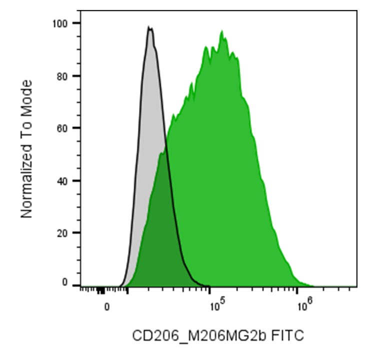
| Cat # | Size | Price | Quantity | |
|---|---|---|---|---|
| 205705 | 25 µg | $70 | ||
| 205706 | 100 µg | $160 |
| Clone | M206MG2b |
|---|---|
| Application | Flow Cytometry |
| Reactivity | Mouse |
| Format | FITC |
| Target Name | CD206, MMR, macrophage mannose receptor, MR (mannose receptor), MRC1 |
| Isotype | Mouse IgG2b |
| Antibody Type | Monoclonal |
| Regulatory Status | RUO |
| Formulation | Phosphate-buffered solution, pH 7.2, containing 0.09% sodium azide and 0.2% (w/v) BSA |
| Protein Concentration | 0.2 mg/mL |
| Storage & Handling | The antibody solution should be stored undiluted between 2°C and 8°C, and protected from prolonged exposure to light. Do not freeze. |
| Recommended Usage | For flow cytometric staining, it is recommended to use less than 0.5 µg of this reagent per 0.5-1.0 million cells in a 100 µL volume. Optimal reagent performance should be determined by titration for each specific application. FITC has an excitation max at 493 nm and an emission max at 525 nm. |
| Excitation Laser | Blue Laser (488 nm) |
| See All Formats | Clone M206MG2b |
Mouse CD206, also known as the mannose receptor (MRC1), is a type I transmembrane C-type lectin receptor expressed predominantly on macrophages, immature dendritic cells, and selected endothelial cells. It functions as a pattern-recognition and scavenger receptor, contributing to innate immune surveillance, endocytosis, phagocytosis, and clearance of endogenous and microbial glycoproteins. CD206 also facilitates antigen uptake and trafficking to intracellular compartments, thereby potentially influencing antigen processing and presentation. The mouse Mrc1 gene encodes a protein that is orthologous to human MRC1/CD206.
CD206 is a large glycoprotein containing a short cytoplasmic tail, a single transmembrane domain, and a large extracellular region. The extracellular portion comprises an N-terminal cysteine-rich (CR) domain, a fibronectin type II (FNII) domain, and eight C-type lectin-like domains (CTLDs). CTLD4 and CTLD5 provide most of the carbohydrate-binding activity. CD206 recognizes glycans containing terminal mannose, fucose, and N-acetylglucosamine (GlcNAc), generally through calcium-dependent interactions. The CR domain additionally recognizes sulfated glycans, while the FNII domain can bind collagen-related structures.
CD206 participates in host defense by recognizing mannose-rich structures on microorganisms, including fungi, mycobacteria, and certain viruses. However, some pathogens can exploit CD206-mediated uptake to enter macrophages or alter immune responses. Dysregulated CD206-positive macrophage activity has also been associated with chronic inflammation, tissue remodeling, fibrosis, infection, and tumor-associated macrophage biology. Mouse studies further indicate that MRC1 influences systemic inflammatory responses and susceptibility to severe inflammatory conditions such as sepsis.
Therapeutically, CD206 is attractive primarily as a macrophage-targeting receptor rather than simply a receptor to block. Mannosylated nanoparticles, proteins, antibodies, and drug-delivery systems can exploit CD206-mediated endocytosis to selectively deliver therapeutic or imaging cargo to CD206-positive macrophages. This strategy is being investigated in cancer, infectious diseases, inflammatory disorders, and lysosomal storage diseases. CD206 therefore represents both a useful macrophage biomarker and a promising target for targeted drug delivery.
FITC Mouse IgG2b Isotype Control Antibody
FITC Anti-Mouse CD206 (MMR) Antibody TDS
Have a product or application question? Consult our FAQs or contact us.