| Cat # | Size | Price | Quantity | |
|---|---|---|---|---|
| 801001 | 25 ug | $145 | ||
| 801002 | 100 ug | $295 |
| Application | ELISA, BLI |
|---|---|
| Format | Liquid, Purified |
| Expression Host | HEK293 |
| Target Name | PD-L1, CD274, B7-H1, PDCD1L1, PDCD1LG1, |
| Species | Human |
| Sources | Recombinant Human PD-L1 (Phe19-Thr239) with C-terminus Fc-Avi tag is expressed in HEK293 cells. |
| Accession Number | Q9NZQ7 |
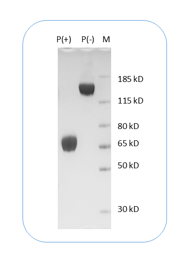
| Molecular Weight | The protein has a predicted molecular weight of 54 kDa and migrates at approximately 70 kDa on SDS-PAGE under DTT-reducing conditions. |
| Affinity Tag | C-Fc-Avi |
| Purity | >95% based on SDS-PAGE under reducing condition |
| Regulatory Status | RUO |
| Formulation | 1xPBS buffer, pH7.4, 0.22 µm filtered |
| Endotoxin level | Not tested |
| Protein Concentration | 25µg size is bottled at 0.2mg/mL concentration. 100 µg size is supplied at a lot-specific concentration. |
| Storage and Handling | Briefly centrifuge the vial upon receipt. An unopened vial can be stored at 4°C for up to 2 weeks, or at -20°C or below for up to six months. The protein may be further diluted to 0.1 mg/mL using 0.22 µm-filtered PBS buffer (pH 7.4). For long-term storage, the diluted stock solution should be aliquoted and stored at ≤ –70°C to minimize freeze-thaw cycles. If additional dilution is required, carrier proteins such as FBS or BSA should be added to maintain protein stability. |
| Research Areas | Activated T cells, Activated B cells, Monocytes, Macrophages, Dendritic cells, NK cells, Immune Checkpoints, Cancer Immunology |
Programmed death-ligand 1 (PD-L1), also known as CD274 or B7-H1, is a transmembrane protein that plays a pivotal role in immune regulation by modulating T cell activity. PD-L1 is expressed on a wide range of cells, including antigen-presenting cells, epithelial cells, and many tumor cells. Its primary function is to bind to its receptor, programmed cell death protein 1 (PD-1), located on activated T cells. This interaction delivers an inhibitory signal that reduces T cell proliferation, cytokine production, and cytotoxicity, thereby maintaining immune homeostasis and preventing autoimmunity. However, in pathological contexts such as cancer, PD-L1 expression allows tumor cells to evade immune attack, creating an immunosuppressive microenvironment.
Structurally, PD-L1 is a type I transmembrane glycoprotein belonging to the B7 family of immune checkpoint molecules. The extracellular domain comprises two immunoglobulin-like regions—an IgV-like domain responsible for PD-1 binding and an IgC-like domain that stabilizes the molecule. The protein also contains a single transmembrane helix and a short cytoplasmic tail that lacks classical signaling motifs but may interact with intracellular partners influencing its stability and localization. The PD-L1–PD-1 complex adopts a well-characterized interface where the IgV domains of both molecules interact in a way that blocks T cell receptor-mediated activation signaling.
The main ligands of PD-L1 are PD-1 and CD80 (B7-1). While PD-1 engagement results in T cell inhibition, interaction with CD80 may yield bidirectional signaling effects depending on the cellular context. PD-L1 can be induced by inflammatory cytokines such as interferon-gamma (IFN-γ), linking innate immune responses to immune checkpoint modulation.
PD-L1 plays a major role in numerous diseases. Overexpression of PD-L1 is a hallmark of many cancers, including lung, melanoma, renal, and breast cancers, where it contributes to immune escape. Therapeutically, blocking the PD-1/PD-L1 axis with immune checkpoint inhibitors has revolutionized cancer treatment. Drugs such as pembrolizumab, nivolumab, and atezolizumab disrupt this inhibitory pathway, restoring antitumor T cell function. Moreover, PD-L1 is being explored as both a predictive biomarker for immunotherapy response and a target for novel therapies, including bispecific antibodies and CAR-T cells aimed at enhancing immune-mediated tumor clearance.
Human PD-L1 (CD274) Protein (C-Fc-Avi) TDS
Anti-Human PD1 (Camrelizumab Biosimilar), Clone Camrelizumab
Anti-Human PD1 (Cemiplimab Biosimilar), Clone Cemiplimab
Anti-Human PD1 (Nivolumab Biosimilar), Clone Nivolumab
Anti-Human PD1 (Pembrolizumab Biosimilar), Clone Pembrolizumab
Anti-Mouse CD274 (PD-L1) Antibody, Clone 10F.9G2
Biotin Anti-Mouse CD274 (PD-L1) Antibody, Clone 10F.9G2
Anti-Human PD-L1 (Atezolizumab Biosimilar), Clone Atezolizuma
Anti-Human PD-L1 (Avelumab Biosimilar), Clone Avelumab
Human PD-L1 (CD274) Protein (C-His)
Human PD-L1 (CD274) Protein (C-His-Avi)
Have a product or application question? Consult our FAQs or contact us.