| Cat # | Size | Price | Quantity | |
|---|---|---|---|---|
| 813401 | 25 ug | $145 | ||
| 813402 | 100 ug | $295 |
| Application | ELISA, BLI |
|---|---|
| Format | Liquid, Purified |
| Expression Host | CHO |
| Target Name | PDL2, , Butyrophilin B7-DC, CD273, PDCD1 ligand 2 |
| Species | Human |
| Sources | Recombinant Human PD-L2 (Leu20-Pro219) with C-terminus Fc-Avi-tag is expressed in CHO cell. |
| Accession Number | Q9BQ51 |
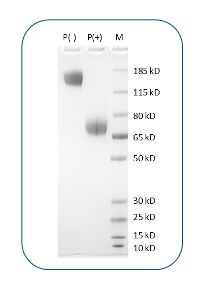
| Molecular Weight | The protein has a predicted molecular weight of 51.2 kDa. Under DTT-reducing conditions, it migrates at approximately 65-75 kDa on SDS-PAGE. |
| Affinity Tag | C-Fc-Avi |
| Purity | >95% based on SDS-PAGE under reducing condition |
| Regulatory Status | RUO |
| Formulation | 1xPBS buffer, pH7.4, 0.22 µm filtered |
| Endotoxin level | Not tested |
| Protein Concentration | 25µg size is bottled at 0.2mg/mL concentration. 100 µg size is supplied at a lot-specific concentration. |
| Storage and Handling | Briefly centrifuge the vial upon receipt. An unopened vial can be stored at 4°C for up to 2 weeks, or at -20°C or below for up to six months. The protein may be further diluted to 0.1 mg/mL using 0.22 µm-filtered PBS buffer (pH 7.4). For long-term storage, the diluted stock solution should be aliquoted and stored at ≤ –70°C to minimize freeze-thaw cycles. If additional dilution is required, carrier proteins such as FBS or BSA should be added to maintain protein stability. |
| Research Areas | Hematopoietic Stem cells, Memory B cells, Asthma, Cancer Marker, Immune Checkpoints, Cancer Immunology |
CD273, also known as programmed death-ligand 2 (PD-L2) or B7-DC, is a transmembrane protein that functions as an immune checkpoint molecule regulating T cell activation and immune responses. CD273 is a member of the B7 family of costimulatory molecules and is primarily expressed on antigen-presenting cells such as dendritic cells, macrophages, and B cells, with expression upregulated during inflammation and immune activation. The protein binds to programmed cell death protein 1 (PD-1) on T cells, delivering inhibitory signals that suppress T cell proliferation, cytokine production, and effector functions. This interaction plays a crucial role in maintaining immune homeostasis, preventing autoimmunity, and resolving inflammatory responses. PD-L2 plays a pivotal role in negative regulation of the adaptive immune response, and unlike its related molecule PD-L1 (CD274), CD273 expression is more restricted and primarily induced under specific inflammatory conditions.
Structurally, CD273 is a type I transmembrane glycoprotein of approximately 25-30 kDa belonging to the immunoglobulin superfamily. The extracellular region contains two immunoglobulin-like domains: an N-terminal IgV-like domain that mediates PD-1 binding and a membrane-proximal IgC-like domain that provides structural support. The protein also contains a single transmembrane domain and a short cytoplasmic tail. Structural studies have revealed the features that distinguish PD-L2 from PD-L1 in their interactions with PD-1. The IgV domain of CD273 interacts with PD-1 through a binding interface similar to that of PD-L1, though CD273 binds PD-1 with approximately 2-6 fold higher affinity than PD-L1. This higher binding affinity allows CD273 to effectively engage PD-1 and deliver coinhibitory signals that modulate T cell responses.
The primary ligand for CD273 is PD-1 (CD279), an inhibitory receptor expressed on activated T cells, B cells, and myeloid cells. PD-1 has two naturally occurring ligands: PD-L1 and PD-L2 (CD273), both identified over a decade ago. The CD273-PD-1 interaction delivers negative regulatory signals that attenuate immune responses. Additionally, CD273 has been reported to bind to repulsive guidance molecule b (RGMb), though the functional significance of this interaction remains under investigation. The binding of CD273 to PD-1 results in recruitment of phosphatases that inhibit T cell receptor signaling pathways, reducing T cell activation and promoting immune tolerance.
In disease contexts, CD273 plays a significant role in cancer immune evasion and is associated with patient prognosis. As an immune checkpoint molecule, PD-L2 was reported to be associated with patient outcomes and plays a pivotal role in cancer cell immune escape. Many tumors upregulate CD273 expression to suppress antitumor T cell responses, contributing to immune evasion. Elevated CD273 expression has been observed in various cancers, including Hodgkin lymphoma, mediastinal B cell lymphoma, and certain solid tumors, where it correlates with immune suppression in the tumor microenvironment. Therapeutically, the evolving landscape of PD-L2 is bringing new light to checkpoint immunotherapy. While most immune checkpoint inhibitors have focused on the PD-1/PD-L1 axis, the PD-1/PD-L2 (CD273) pathway is also being investigated as a therapeutic target. Some anti-PD-1 antibodies such as nivolumab and pembrolizumab block both PD-L1 and PD-L2 binding to PD-1, contributing to their therapeutic efficacy. Additionally, CD273 is being explored as a biomarker for predicting response to immunotherapy, and selective CD273-targeting strategies are under development to modulate immune responses in cancer and autoimmune diseases while potentially minimizing toxicity compared to broader PD-1/PD-L1 blockade.
Human PD-L2 (CD273) Protein (C-Fc-Avi) TDS
Human PD-L2 (CD273) Protein (C-His-Avi)
Biotin Human PD-L2 (CD273) Protein (C-His-Avi)
Human PD-L2 (CD273) Protein (C-His)
PE Human PD-L2 (CD273) Protein (C-His)
APC Human PD-L2 (CD273) Protein (C-His)
Biotin Human PD-L2 (CD273) Protein (C-Fc-Avi)
Human PD-L2 (CD273) Protein (C-Fc)
PE Human PD-L2 (CD273) Protein (C-Fc)
APC Human PD-L2 (CD273) Protein (C-Fc)
Have a product or application question? Consult our FAQs or contact us.