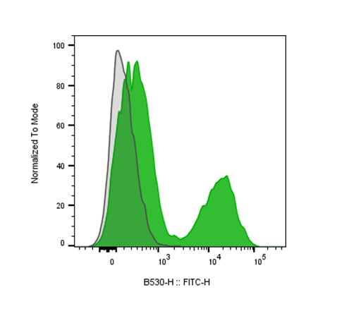
| Cat # | Size | Price | Quantity | |
|---|---|---|---|---|
| 201707 | 25 tests | $80 | ||
| 201708 | 100 tests | $180 |
| Clone | H57-597-R1 |
|---|---|
| Application | Flow Cytometry |
| Reactivity | Mouse |
| Format | iF488 |
| Target Name | TCR β chain, TCR-β |
| Isotype | Rat IgG1 |
| Antibody Type | Monoclonal |
| Regulatory Status | RUO |
| Formulation | Phosphate-buffered solution, pH 7.2, containing 0.09% sodium azide and 0.2% (w/v) BSA |
| Protein Concentration | Supplied at a lot-specific concentration. |
| Storage&Handling | The antibody solution should be stored undiluted between 2°C and 8°C, and protected from prolonged exposure to light. Do not freeze. |
| Recommended Usage | For flow cytometric staining, it is recommended to use 5 µL of this reagent per 0.5-1.0 million cells in a 100 µL volume. Optimal reagent performance should be determined by titration for each specific application. iF488 has an excitation max at 491 nm and an emission max at 516 nm. |
| Excitation Laser | Blue Laser (488 nm) |
| RRID | AB_3739062 |
| Research Areas | Antigen presentation, T cells, T cell Activation |
| See All Formats | Clone H57-597-R1 |
The mouse T cell receptor (TCR) β chain is a critical component of the heterodimeric αβ TCR complex expressed on most mature T lymphocytes. Together with the TCR α chain, it confers antigen specificity to T cells, enabling the recognition of peptide fragments presented by major histocompatibility complex (MHC) molecules on antigen-presenting cells. This recognition event is essential for initiating adaptive immune responses, including T cell activation, proliferation, and differentiation into effector and memory subsets.
Structurally, the TCR β chain is composed of variable (V), diversity (D), joining (J), and constant (C) gene segments that undergo somatic recombination during T cell development in the thymus. This recombination process generates the highly variable complementarity-determining region 3 (CDR3), which contributes most significantly to antigen specificity. The β chain pairs non-covalently with the TCR α chain, forming the antigen-binding site. Each chain contains two extracellular immunoglobulin-like domains, a variable domain involved in antigen binding and a constant domain that stabilizes structure, along with a transmembrane segment and a short cytoplasmic tail. The TCR complex also associates with CD3 signaling molecules (CD3γ, CD3δ, CD3ε, and CD3ζ), which transduce activation signals through immunoreceptor tyrosine-based activation motifs (ITAMs).
The ligands for the mouse TCR β chain are peptide antigens bound to MHC class I or II molecules. Engagement of the αβ TCR with these peptide-MHC complexes triggers receptor conformational changes that initiate intracellular signaling cascades involving kinases such as Lck and ZAP-70, leading to T cell activation and effector function.
In disease, alterations in TCR β chain expression or repertoire diversity can contribute to immune dysfunction. Restricted TCR β repertoires are associated with autoimmune diseases, such as experimental autoimmune encephalomyelitis (EAE, a model for multiple sclerosis), and with impaired immune defense in infections or cancer. Somatic mutations or skewed TCR β usage have also been observed in T cell lymphomas and leukemia.
The mouse TCR β chain has significant therapeutic and experimental relevance. It serves as a model for studying clonal selection, antigen recognition, and tolerance mechanisms. In immunotherapy, manipulation of TCR β sequence diversity underpins TCR-engineered T cell strategies for cancer and infection control. Furthermore, analyzing mouse TCR β repertoires provides insights into vaccine efficacy, autoimmune mechanisms, and immune reconstitution following bone marrow transplantation.
iF488 Rat IgG1 Isotype Control Antibody
iF488 Anti-Mouse TCR β chain Antibody TDS
APC Anti-Mouse TCR β chain Antibody, Clone H57-597-M2a
In Vivo Star Anti-Mouse TCR beta Antibody, Clone H57-597-m2a
Have a product or application question? Consult our FAQs or contact us.