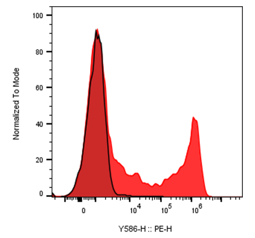
| Cat # | Size | Price | Quantity | |
|---|---|---|---|---|
| 105921 | 25 tests | $105 | ||
| 105922 | 100 tests | $225 |
| Clone | 3G8 |
|---|---|
| Application | Flow Cytometry |
| Reactivity | Human |
| Format | iF560 |
| Target Name | CD16, FcγRIII |
| Isotype | Mouse IgG1 |
| Antibody Type | Monoclonal |
| Regulatory Status | RUO |
| Formulation | Phosphate-buffered solution, pH 7.2, containing 0.09% sodium azide and 0.2% (w/v) BSA |
| Protein Concentration | Supplied at a lot-specific concentration. |
| Storage & Handling | The antibody solution should be stored undiluted between 2°C and 8°C, and protected from prolonged exposure to light. Do not freeze. |
| Recommended Usage | For flow cytometric staining, it is recommended to use 5 µL of this reagent per 0.5-1.0 million cells in a 100 µL volume. Optimal reagent performance should be determined by titration for each specific application. iF560 has an excitation max at 560 nm and an emission max at 571 nm. |
| Excitation Laser | Green/Yellow laser (532/561nm) |
| See All Formats | Clone 3G8 |
CD16, also known as Fc gamma receptor III (FcγRIII), is a low-affinity receptor for the Fc region of immunoglobulin G (IgG) and plays a pivotal role in antibody-mediated immune responses. It is expressed primarily on natural killer (NK) cells, neutrophils, monocytes, and macrophages, with expression patterns and function varying by cell type. In humans, CD16 exists in two closely related forms encoded by distinct genes: CD16a (FcγRIIIA) and CD16b (FcγRIIIB).
Structurally, CD16 is a type I transmembrane glycoprotein composed of two extracellular immunoglobulin-like domains responsible for IgG binding. CD16a is a transmembrane receptor expressed on NK cells and some myeloid cells, where it associates with signaling adaptor proteins containing immunoreceptor tyrosine-based activation motifs (ITAMs), such as FcεRIγ and CD3ζ. In contrast, CD16b is attached to the cell surface via a glycosylphosphatidylinositol (GPI) anchor and is expressed almost exclusively on neutrophils, lacking direct intracellular signaling capacity.
Functionally, CD16 mediates antibody-dependent cellular cytotoxicity (ADCC) and phagocytosis. Upon binding to IgG-opsonized target cells, CD16a on NK cells triggers activation signals that lead to the release of cytotoxic granules containing perforin and granzymes, resulting in target cell death. On myeloid cells, CD16 engagement promotes phagocytosis, oxidative burst, and cytokine release, contributing to pathogen clearance and inflammation. CD16 preferentially binds IgG1 and IgG3 subclasses, which are commonly elicited during effective immune responses.
CD16 plays important roles in both protective immunity and disease. Genetic polymorphisms in FCGR3A influence IgG binding affinity and have been associated with susceptibility to infections, autoimmune diseases, and cancer outcomes. Excessive or dysregulated CD16 activation contributes to inflammatory and autoimmune conditions such as rheumatoid arthritis and systemic lupus erythematosus, where immune complexes drive tissue damage. In cancer, CD16 expression and function on NK cells are critical determinants of immune surveillance and therapeutic efficacy.
In therapeutics, CD16 is central to the mechanism of action of many antibody-based drugs. Therapeutic monoclonal antibodies used in oncology, such as those targeting tumor antigens, rely on CD16-mediated ADCC for clinical activity. Engineering antibodies with enhanced Fc affinity for CD16 or developing CD16-engaging bispecific antibodies are active areas of drug development. Additionally, adoptive NK cell therapies often aim to optimize CD16 expression and signaling, highlighting CD16 as a key bridge between antibody recognition and cellular immune effector function.
iF560 Mouse IgG1 Isotype Control Antibody
iF560 Anti-Human CD16 Antibody TDS
Have a product or application question? Consult our FAQs or contact us.