| Cat # | Size | Price | Quantity | |
|---|---|---|---|---|
| 116701 | 25 tests | $120 | ||
| 116702 | 100 tests | $250 |
| Clone | Gemtuzumab |
|---|---|
| Application | Flow Cytometry |
| Reactivity | Human |
| Format | iF647 |
| Target Name | CD33, Siglec-3, gp67, p67, Siglec3 |
| Isotype | Human IgG4 |
| Antibody Type | Monoclonal |
| Regulatory Status | RUO |
| Formulation | Phosphate-buffered solution, pH 7.2, containing 0.09% sodium azide and 0.2% (w/v) BSA |
| Protein Concentration | Supplied at a lot-specific concentration. |
| Storage&Handling | The antibody solution should be stored undiluted between 2°C and 8°C, and protected from prolonged exposure to light. Do not freeze. |
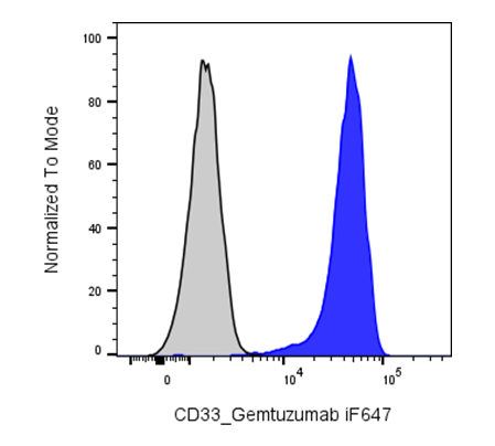
| Recommended Usage | For flow cytometric staining, it is recommended to use 5 µL of this reagent per 0.5-1.0 million cells in a 100 µL volume. Optimal reagent performance should be determined by titration for each specific application. iF647 has an excitation max at 656 nm and an emission max at 670 nm. |
| Excitation Laser | Red Laser (633 nm) |
| Research Areas | Monocytes, Macrophages, Dendritic cells, Innate Immunity, Cancer Marker |
| See All Formats | Clone Gemtuzumab |
Human CD33 is a sialic acid–binding immunoglobulin-like lectin (Siglec) expressed predominantly on myeloid lineage cells, including hematopoietic progenitors, monocytes, and myeloid blasts in acute myeloid leukemia (AML). Structurally, CD33 is a type I transmembrane glycoprotein composed of an N-terminal V-set Ig-like domain that mediates ligand recognition, a C2-set Ig-like domain, a single transmembrane region, and a cytoplasmic tail containing immunoreceptor tyrosine-based inhibitory motifs (ITIMs). These ITIMs recruit phosphatases such as SHP-1 and SHP-2 to transmit inhibitory signaling that dampens cellular activation.
CD33 recognizes sialylated glycans on host cell surfaces as its endogenous ligands, enabling it to function as a “self-recognition” receptor that helps regulate myeloid cell activation, cytokine release, and phagocytosis. Through these interactions, CD33 contributes to the maintenance of immune homeostasis and prevention of excessive inflammation. However, this inhibitory signaling axis can be co-opted in disease states.
In acute myeloid leukemia, CD33 is highly expressed on leukemic blasts, making it a clinically important biomarker and therapeutic target. Its expression is generally absent or low on non-hematopoietic tissues, which improves its suitability for targeted therapy. Overexpression of CD33 in AML is associated with disease burden and progression, and it is frequently used for diagnosis and stratification.
Gemtuzumab is a humanized monoclonal antibody directed against CD33, and it is conjugated to the cytotoxic agent calicheamicin. Upon binding CD33 on AML cells, the antibody–drug conjugate is internalized, releasing calicheamicin intracellularly to induce DNA double-strand breaks and apoptosis. Clinically, gemtuzumab ozogamicin has been used in AML therapy, either as monotherapy or in combination regimens, improving outcomes in selected patient populations by selectively targeting CD33-positive leukemic cells while sparing most normal tissues.
iF647 Human IgG4 Isotype Control Antibody
iF647 Anti-Human CD33 Antibody TDS
Have a product or application question? Consult our FAQs or contact us.