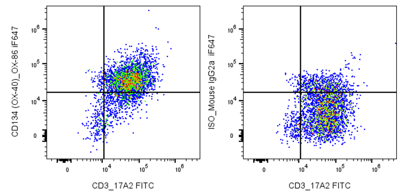
| Cat # | Size | Price | Quantity | |
|---|---|---|---|---|
| 207903 | 25 µg | $95 | ||
| 207904 | 100 µg | $245 |
| Clone | OX-86 |
|---|---|
| Application | Flow Cytometry |
| Reactivity | Mouse |
| Format | iF647 |
| Target Name | CD134, OX-40, OX40, TNFRSF4, ACT35 |
| Isotype | Rat IgG1 |
| Antibody Type | Monoclonal |
| Regulatory Status | RUO |
| Formulation | Phosphate-buffered solution, pH 7.2, containing 0.09% sodium azide and 0.2% (w/v) BSA |
| Protein Concentration | 0.2 mg/mL |
| Storage & Handling | The antibody solution should be stored undiluted between 2°C and 8°C, and protected from prolonged exposure to light. Do not freeze. |
| Recommended Usage | For flow cytometric staining, it is recommended to use 5 µL of this reagent per 0.5-1.0 million cells in a 100 µL volume. Optimal reagent performance should be determined by titration for each specific application. iF647 has an excitation max at 656 nm and an emission max at 670 nm. |
| Excitation Laser | Red Laser (633 nm) |
| See All Formats | Clone OX-86 |
Mouse CD134, also known as OX40 or TNFRSF4, is a member of the tumor necrosis factor receptor (TNFR) superfamily and an important costimulatory receptor on T cells. OX40 is induced following T-cell receptor activation and is expressed predominantly on activated CD4+ and CD8+ T cells. In mice, OX40 is also constitutively expressed on certain regulatory T cells (Tregs). OX40 signaling promotes T-cell proliferation, survival, differentiation, cytokine production, and formation of memory T cells, thereby strengthening and sustaining antigen-specific immune responses.
Mouse OX40 is a type I transmembrane glycoprotein belonging to the TNFR superfamily. It contains an extracellular region with characteristic cysteine-rich domains, a single transmembrane segment, and a cytoplasmic signaling tail. The principal and currently established ligand is OX40 ligand (OX40L), also known as CD252 or TNFSF4. OX40L is a type II transmembrane TNF-superfamily protein expressed primarily by activated antigen-presenting cells, including dendritic cells, macrophages, and B cells. OX40L forms a trimer and engages OX40 to initiate downstream signaling through pathways including NF-κB and PI3K/Akt.
Excessive OX40 signaling can contribute to chronic inflammation and autoimmune disease. In mouse models, the OX40–OX40L pathway has been implicated in experimental autoimmune encephalomyelitis, asthma, colitis, diabetes, arthritis, graft-versus-host disease, and transplant rejection. Blocking OX40 or OX40L can substantially reduce disease severity in several of these models, supporting the pathway as a potential therapeutic target.
Therapeutically, OX40 can be targeted in two opposing ways. Agonistic anti-OX40 antibodies or OX40L-based agonists can enhance T-cell activation and antitumor immunity, making OX40 an attractive target for cancer immunotherapy and vaccine development. Conversely, antagonistic antibodies or OX40L blockade may suppress pathogenic T-cell responses in autoimmune and inflammatory diseases. OX40 agonists, including combinations with immune-checkpoint inhibitors, continue to be investigated clinically, although antitumor efficacy has so far been variable.
iF647 Rat IgG1 Isotype Control Antibody
iF647 Anti-Mouse CD134 (OX-40) Antibody TDS
Have a product or application question? Consult our FAQs or contact us.