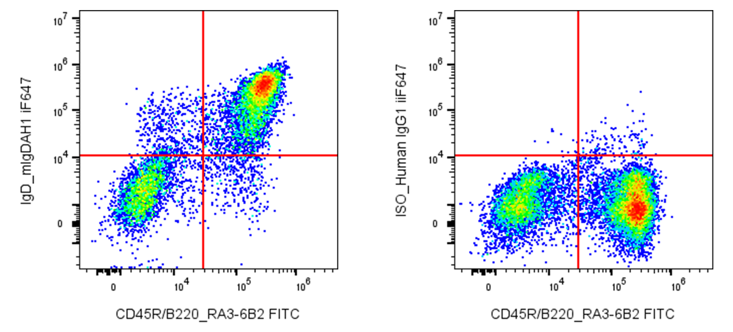
| Cat # | Size | Price | Quantity | |
|---|---|---|---|---|
| 208703 | 25 µg | $95 | ||
| 208704 | 100 µg | $220 |
| Clone | mIgDAH1 |
|---|---|
| Application | Flow Cytometry |
| Reactivity | Mouse |
| Format | iF647 |
| Target Name | IgD, Immunoglobulin D |
| Isotype | Human IgG1 |
| Antibody Type | Monoclonal |
| Regulatory Status | RUO |
| Formulation | Phosphate-buffered solution, pH 7.2, containing 0.09% sodium azide and 0.2% (w/v) BSA |
| Protein Concentration | 0.2 mg/mL |
| Storage & Handling | The antibody solution should be stored undiluted between 2°C and 8°C, and protected from prolonged exposure to light. Do not freeze. |
| Recommended Usage | For flow cytometric staining, it is recommended to use less than 0.25 µg of this reagent per 0.5-1.0 million cells in a 100 µL volume. Optimal reagent performance should be determined by titration for each specific application. iF647 has an excitation max at 656 nm and an emission max at 670 nm. |
| Excitation Laser | Red Laser (633 nm) |
| See All Formats | Clone mIgDAH1 |
Immunoglobulin D (IgD) is one of the five major classes of immunoglobulins in humans and plays a role in B-cell development and immune regulation. It is expressed primarily on the surface of mature, naïve B lymphocytes, where it is co-expressed with immunoglobulin M (IgM) and functions as part of the B-cell receptor (BCR) complex. When an antigen binds and cross-links surface IgD, it can initiate intracellular signaling that influences B-cell activation, maturation, and differentiation. A smaller amount of IgD is also found in soluble form in the blood and other secretions, although its functions are less completely understood than those of IgG or IgM.
IgD is a glycoprotein composed of two identical heavy chains of the delta (δ) type and two identical light chains, either kappa or lambda. Each heavy chain contains variable and constant regions, with the variable regions contributing to antigen recognition. The constant region of the δ heavy chain includes three immunoglobulin-like domains and a hinge region that provides flexibility. Membrane-bound IgD associates with the signaling proteins Igα (CD79A) and Igβ (CD79B), which transmit signals into the B cell. Unlike some immunoglobulins, IgD does not have a single universal ligand; its antigen-binding sites recognize diverse antigens according to the variable-region sequence. IgD has also been reported to interact with basophils and other innate immune cells, although the physiological significance of some proposed interactions remains under investigation.
IgD is important for normal B-cell biology, and abnormal IgD production or regulation can occur in disease. Elevated serum IgD may be observed in certain inflammatory conditions, infections, and rare disorders such as hyper-IgD syndrome, which is commonly associated with mevalonate kinase deficiency. IgD monoclonal proteins can also occur in rare cases of IgD multiple myeloma. However, an elevated IgD level alone is not diagnostic of a specific disease.
Therapeutically, IgD is useful as a marker for studying and identifying naïve B cells in immunology research and flow cytometry. Targeting IgD-positive B cells or modulating IgD-mediated signaling may offer research avenues for selected immune disorders, but IgD itself is not currently a widely established therapeutic target. Further investigation is needed to clarify its functions and clinical potential.
iF647 Human IgG1 Isotype Control Antibody
iF647 Anti-mouse IgD Antibody TDS
Have a product or application question? Consult our FAQs or contact us.