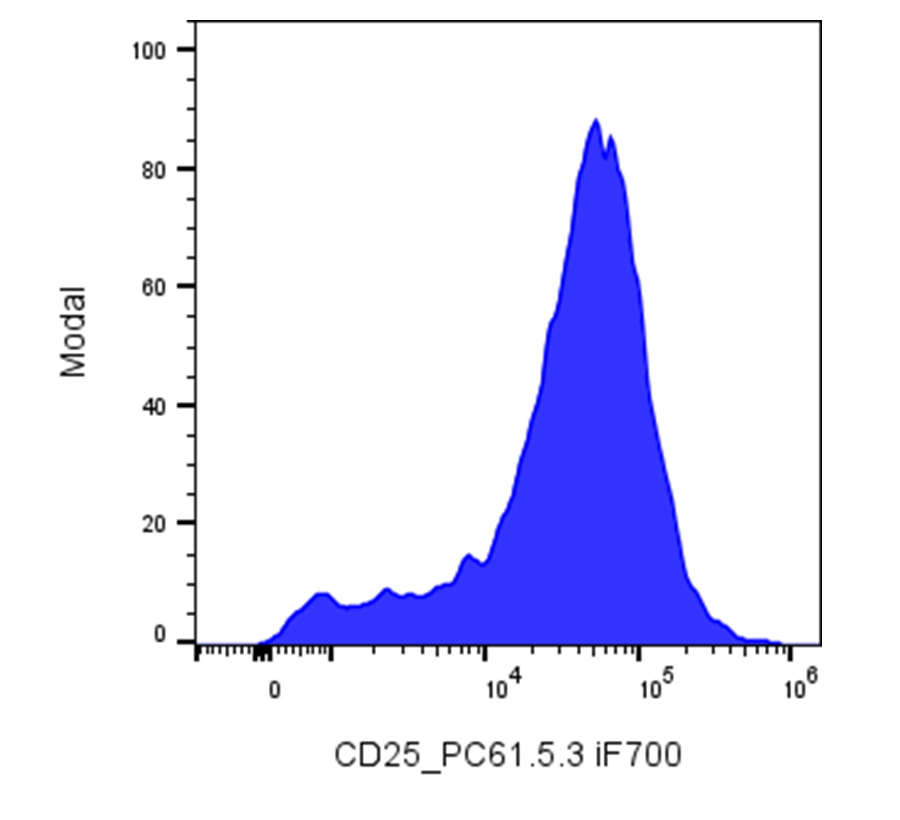
| Cat # | Size | Price | Quantity | |
|---|---|---|---|---|
| 206709 | 25 µg | $90 | ||
| 206710 | 100 µg | $195 |
| Clone | PC61.5.3 |
|---|---|
| Application | Flow Cytometry |
| Reactivity | Mouse |
| Format | iF700 |
| Target Name | IL-2 receptor α chain, Low affinity IL-2R, IL-2Rα chain |
| Isotype | Rat IgG1 |
| Antibody Type | Monoclonal |
| Regulatory Status | RUO |
| Formulation | Phosphate-buffered solution, pH 7.2, containing 0.09% sodium azide and 0.2% (w/v) BSA |
| Protein Concentration | 0.2 mg/mL |
| Storage & Handling | The antibody solution should be stored undiluted between 2°C and 8°C, and protected from prolonged exposure to light. Do not freeze. |
| Recommended Usage | For flow cytometric staining, it is recommended to use less than 0.2 µg of this reagent per 0.5-1.0 million cells in a 100 µL volume. Optimal reagent performance should be determined by titration for each specific application. iF647 has an excitation max at 656 nm and an emission max at 670 nm. |
| Excitation Laser | Red Laser (633 nm) |
| See All Formats | Clone PC61.5.3 |
CD25, also known as the interleukin-2 receptor alpha chain (IL-2Rα), is a transmembrane glycoprotein that plays a central role in regulating immune responses. It functions as part of the interleukin-2 (IL-2) receptor complex, which is essential for T cell proliferation, survival, and differentiation. CD25 itself has low affinity for IL-2 when expressed alone, but when combined with IL-2 receptor beta (CD122) and the common gamma chain (CD132), it forms the high-affinity IL-2 receptor complex capable of effective signal transduction.
Structurally, CD25 is a single-pass type I membrane protein composed of an extracellular domain of approximately 219 amino acids responsible for IL-2 binding, a hydrophobic transmembrane segment, and a short cytoplasmic tail that lacks intrinsic signaling domains. The extracellular region is heavily glycosylated, which stabilizes its conformation and facilitates ligand interaction. Because the alpha chain alone is not signaling-competent, it acts primarily to increase the receptor complex’s affinity for IL-2 and to expand the range of cells responsive to low cytokine concentrations.
CD25’s main ligand, IL-2, is a cytokine crucial for T lymphocyte expansion and immune tolerance. Engagement of IL-2 with the high-affinity receptor triggers the JAK-STAT signaling pathway, leading to cell proliferation, differentiation, and regulatory T cell (Treg) function. CD25 is constitutively expressed on Tregs and upregulated on activated CD4+ and CD8+ T cells, making it a marker of immune activation as well as immune regulation.
Aberrant CD25 expression or IL-2 signaling contributes to immune dysregulation and disease. In autoimmune disorders such as multiple sclerosis and type 1 diabetes, alterations in the IL-2/CD25 axis impair Treg function and tolerance mechanisms. Elevated CD25 expression is also found in certain malignancies, particularly adult T-cell leukemia/lymphoma and Hodgkin lymphoma, where it may serve as a biomarker of malignant proliferation. Moreover, soluble CD25, released from cell surfaces, can act as a decoy receptor, modulating IL-2 availability and contributing to immune suppression in cancer and chronic inflammation.
Therapeutically, CD25 is a prominent target for immune modulation. Monoclonal antibodies such as basiliximab and daclizumab have been developed to block IL-2 binding, preventing T cell activation and mitigating graft rejection in organ transplantation. Conversely, IL-2 or CD25-targeted therapies that enhance regulatory T cell function are being explored to treat autoimmune diseases and promote immune tolerance. Thus, CD25 remains a critical immunological node, balancing activation and regulation within the immune system.
iF700 Rat IgG1 Isotype Control
iF700 Anti-Mouse CD25 Antibody TDS
Have a product or application question? Consult our FAQs or contact us.