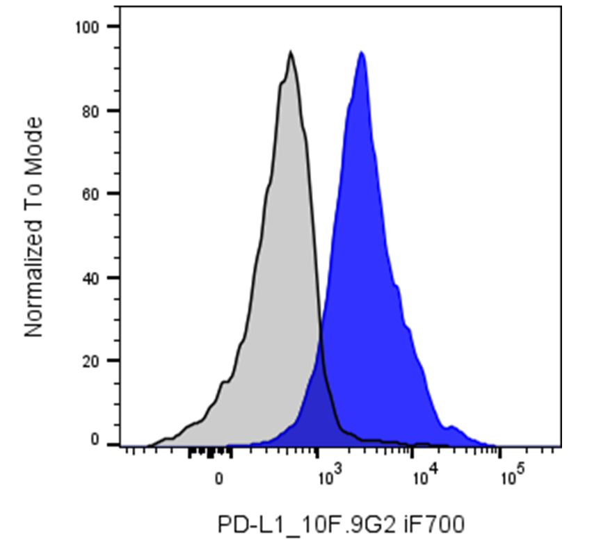
| Cat # | Size | Price | Quantity | |
|---|---|---|---|---|
| 201317 | 25 μg | $120 | ||
| 201318 | 100 μg | $280 |
| Clone | 10F.9G2 |
|---|---|
| Application | Flow Cytometry |
| Reactivity | Mouse |
| Format | iF700 |
| Target Name | CD274, PD-L1, B7-H1 |
| Isotype | Rat IgG2b |
| Antibody Type | Monoclonal |
| Regulatory Status | RUO |
| Formulation | Phosphate-buffered solution, pH 7.2, containing 0.09% sodium azide and 0.2% (w/v) BSA |
| Protein Concentration | 0.2 mg/mL |
| Storage & Handling | The antibody solution should be stored undiluted between 2°C and 8°C, and protected from prolonged exposure to light. Do not freeze. |
| Recommended Usage | For flow cytometric staining, it is recommended to use less than 0.25 µg of this reagent per 0.5-1.0 million cells in a 100 µL volume. Optimal reagent performance should be determined by titration for each specific application. iF700 has an excitation max at 690 nm and an emission max at 710 nm. |
| Excitation Laser | Red Laser (633 nm) |
| See All Formats | Clone 10F.9G2 |
PD-L1 (Programmed Death-Ligand 1), also known as CD274 or B7-H1, is a 40 kDa type I transmembrane protein belonging to the B7 family within the immunoglobulin receptor superfamily. The protein contains immunoglobulin V-like and C-like domains and is expressed by a wide range of hematopoietic and non-hematopoietic cells, including T cells, B cells, NK cells, dendritic cells, monocytes, endothelial cells, and various tumor cells. PD-L1 serves as a ligand for PD-1 (CD279) and plays a critical role in immune regulation by inhibiting T-cell activation, proliferation, and cytokine production upon engagement with PD-1. This interaction maintains immune homeostasis during infection or inflammation, preventing autoimmunity. However, in tumor microenvironments, PD-L1 expression enables immune evasion by suppressing cytotoxic T-cell function, contributing to tumor progression. PD-L1 expression is considered prognostic in several malignancies, including colon cancer and renal cell carcinoma. Alternative splicing results in multiple transcript variants. The PD-1/PD-L1 axis is a major target in cancer immunotherapy.
iF700 Rat IgG2b Isotype Control
iF700 Anti-Mouse CD274 (PD-L1) Antibody TDS
Have a product or application question? Consult our FAQs or contact us.