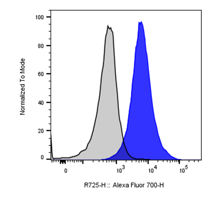
| Cat # | Size | Price | Quantity | |
|---|---|---|---|---|
| 200615 | 25 tests | $75 | ||
| 200616 | 100 tests | $170 |
| Clone | D7 |
|---|---|
| Application | Flow Cytometry |
| Reactivity | Mouse |
| Format | iF700 |
| Target Name | Ly-6A/E, Sca-1 |
| Isotype | Rat IgG2a |
| Antibody Type | Monoclonal |
| Regulatory Status | RUO |
| Formulation | Phosphate-buffered solution, pH 7.2, containing 0.09% sodium azide and 0.2% (w/v) BSA |
| Protein Concentration | Supplied at a lot-specific concentration. |
| Storage&Handling | The antibody solution should be stored undiluted between 2°C and 8°C, and protected from prolonged exposure to light. Do not freeze. |
| Recommended Usage | For flow cytometric staining, it is recommended to use 5 uL of this reagent per 0.5-1.0 million cells in a 100 µL volume. Optimal reagent performance should be determined by titration for each specific application. iF700 has an excitation max at 690 nm and an emission max at 710 nm. |
| Excitation Laser | Red Laser (633 nm) |
| Research Areas | Hematopoietic Stem cells, Mesenchymal Stem cells, Inflammation, Angiogenesis, Cancer Immunology |
| See All Formats | Clone D7 |
Ly-6A/E, commonly known as stem cell antigen-1 (Sca-1), is a glycosylphosphatidylinositol (GPI)-anchored cell surface protein that serves as an important marker for murine hematopoietic stem cells and other progenitor populations. It belongs to the Ly-6/uPAR superfamily, which consists of small, cysteine-rich proteins involved in cell signaling, adhesion, and immune regulation. Functionally, Sca-1 contributes to stem cell maintenance, tissue regeneration, and immune responses, making it a key molecule for studying stem cell biology and immunoregulation in mice.
Structurally, Ly-6A/E is a relatively small protein of about 18-20 kDa, characterized by a conserved “three-finger” Ly-6 domain stabilized by multiple intramolecular disulfide bonds. It is tethered to the cell membrane via a GPI anchor rather than a transmembrane domain, allowing lateral mobility within the lipid bilayer and association with signaling complexes. Sca-1 lacks intrinsic signaling motifs but modulates cell function through its interactions with other membrane-associated proteins, influencing downstream signaling cascades involving Src-family kinases and MAPK pathways.
While the precise ligands of Sca-1 remain not fully defined, its activity is thought to depend on interactions with lipid raft-associated coreceptors and possibly extracellular matrix components. Sca-1 expression is dynamically regulated by cytokines and growth factors, including interferons, which enhance its expression in both stem and immune cells. Sca-1 plays multiple physiological roles. In the hematopoietic system, it marks multipotent progenitors within the bone marrow and contributes to self-renewal and differentiation capacity. In non-hematopoietic tissues, such as skeletal muscle and the heart, Sca-1 marks resident stem or progenitor cells involved in tissue repair following injury. Within the immune system, it is expressed on activated T cells and influences T cell activation and cytokine production.
In disease, altered Sca-1 expression or function has been associated with impaired tissue regeneration, inflammatory conditions, and tumor progression. Sca-1-positive cells have been identified in various cancers, where they may contribute to tumor initiation, metastasis, and resistance to therapy, acting as markers of tumor-propagating cell populations.
Therapeutically, Ly-6A/E serves as a valuable marker for isolating stem and progenitor cells for transplantation and regenerative medicine. Understanding its regulatory role in stem cell signaling could improve strategies for tissue repair and immunotherapy. Additionally, Sca-1-associated pathways may offer targets for modulating cancer stem cell behavior and enhancing therapeutic response in experimental disease models.
iF700 Rat IgG2a Isotype Control
iF700 Anti-Mouse Ly-6A/E Antibody TDS
Have a product or application question? Consult our FAQs or contact us.