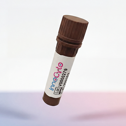
| Cat # | Size | Price | Quantity | |
|---|---|---|---|---|
| 509101 | 1 mg | $160 | ||
| 509102 | 5 mg | $400 | ||
| 509103 | 25 mg | $1100 |
| Clone | 145-2C11-m2a |
|---|---|
| Application | ELISA, WB, Flow cytometry, IHC, ICC, animal model study |
| Host Species | CHO cells |
| Reactivity | Mouse |
| Format | Liquid |
| Target Name | CD3, T3, CD3ε |
| Product Description | In vivo Grade Recombinant Anti-mouse CD3e Monoclonal Antibody |
| Isotype | Mouse IgG2a Kappa |
| Antibody Type | Recombinant |
| Regulatory Status | RUO |
| Purity | >95% by reducing SDS-PAGE |
| Endotoxin | < 1 EU per 1 mg of the protein by the LAL method. |
| Storage Conditions | 4ºC |
| Grade | In vivo |
| Recommended Usage | This product is suitable for in vivo animal use. Optimal amounts need to be determined empirically for each experiment. |
| RRID | AB_3739365 |
| Research Areas | T cells, T cell Activation, Adaptive Immunity |
| See All Formats | Clone 145-2C11-m2a |
CD3 is a critical component of the T-cell receptor (TCR) complex, essential for T-cell activation and the adaptive immune response. It is present on nearly all mature T lymphocytes and serves as a marker for their identification. Functionally, CD3 transduces activation signals from the TCR upon antigen recognition, triggering cascades that lead to cytokine production, cell proliferation, and differentiation. This process enables T-cells to respond effectively to pathogens and abnormal cells, anchoring immune defense mechanisms.
Structurally, CD3 is not a single protein but a multimeric complex composed of four distinct polypeptide chains: CD3γ, CD3δ, CD3ε, and CD3ζ. These chains associate non-covalently with the antigen-specific α and β chains of the TCR. Each CD3 subunit contains one or more immunoreceptor tyrosine-based activation motifs (ITAMs) in their cytoplasmic tails. Upon TCR engagement with an antigen–major histocompatibility complex (MHC), the ITAMs become phosphorylated, serving as docking sites for signaling molecules such as ZAP-70, which propagate downstream signaling required for full T-cell activation. CD3 itself does not bind directly to antigens; instead, it functions as a signaling adaptor within the TCR complex. Its effective operation relies on interactions between the TCR and peptide-MHC complexes presented by antigen-presenting cells. The physical and functional association between CD3 and the TCR ensures that antigen recognition is tightly coupled to intracellular signaling pathways that determine T-cell fate and function.
Alterations in CD3 expression or signaling are implicated in various immune-related diseases. Mutations in CD3 genes can cause severe combined immunodeficiency (SCID), characterized by defective T-cell development. Dysregulated signaling may also contribute to autoimmune conditions, where inappropriate T-cell activation leads to tissue damage. Additionally, CD3 surface expression levels are sometimes altered in chronic infections and leukemias.
Therapeutically, CD3 is an important target in immunomodulation. Monoclonal antibodies such as muromonab-CD3 (OKT3) were among the first biologics used to prevent organ transplant rejection by depleting or inactivating T-cells. More recently, bispecific T-cell engagers (BiTEs) like blinatumomab exploit CD3 to redirect cytotoxic T-cells toward tumor cells expressing specific antigens, offering potent anti-cancer activity. By harnessing or modulating CD3-mediated signaling, modern immunotherapies continue to expand the therapeutic potential of T-cell-based treatments.
In Vivo Star Anti-Mouse CD3e Antibody TDS
In Vivo Star Anti-Mouse CD3e Antibody, Clone 500A2
Have a product or application question? Consult our FAQs or contact us.