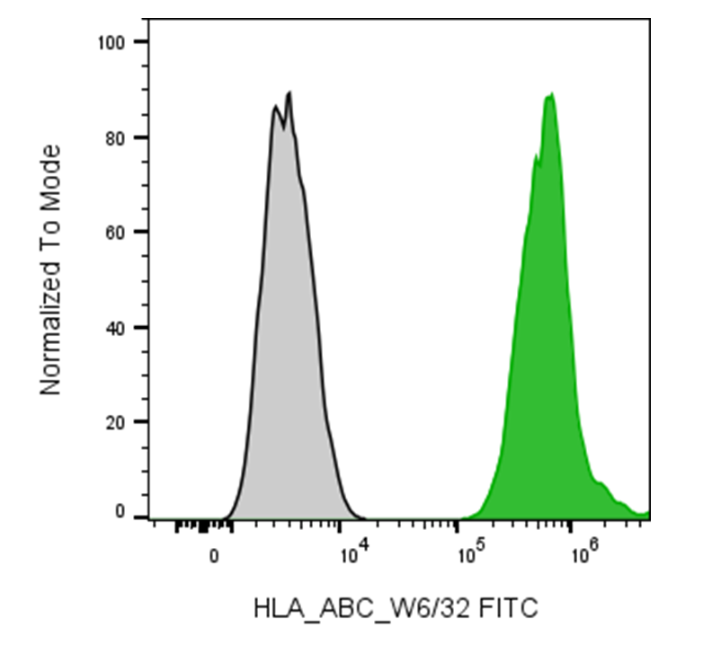
| Cat # | Size | Price | Quantity | |
|---|---|---|---|---|
| 180607 | 25 tests | $125 | ||
| 180608 | 100 tests | $250 |
| Clone | HLACB |
|---|---|
| Application | Flow Cytometry |
| Reactivity | Human |
| Format | FITC |
| Target Name | HLA-ABC, Major Histocompatibility Class I, MHC class I |
| Isotype | Rabbit IgG |
| Antibody Type | Monoclonal |
| Regulatory Status | RUO |
| Formulation | Phosphate-buffered solution, pH 7.2, containing 0.09% sodium azide and 0.2% (w/v) BSA |
| Protein Concentration | Supplied at a lot-specific concentration. |
| Storage & Handling | The antibody solution should be stored undiluted between 2°C and 8°C, and protected from prolonged exposure to light. Do not freeze. |
| Recommended Usage | For flow cytometric staining, it is recommended to use 5 µL of this reagent per 0.5-1.0 million cells in a 100 µL volume. Optimal reagent performance should be determined by titration for each specific application. FITC has an excitation max at 493 nm and an emission max at 525 nm. |
| Excitation Laser | Blue Laser (488 nm) |
| Brand | InnoSite |
| See All Formats | Clone HLACB |
HLA-ABC refers to the classical human leukocyte antigen (HLA) class I molecules, HLA-A, HLA-B, and HLA-C, encoded within the major histocompatibility complex (MHC) on chromosome 6. These molecules are expressed on the surface of nearly all nucleated cells and play a pivotal role in the adaptive immune response. Their main function is to present endogenously derived peptide antigens, typically from intracellular proteins, to cytotoxic CD8⁺ T lymphocytes. This antigen presentation enables the immune system to continuously monitor cell integrity and identify cells infected by viruses or transformed by cancer.
Structurally, HLA class I molecules are heterodimeric complexes consisting of a transmembrane heavy (alpha) chain and a non-covalently associated light chain known as β2-microglobulin. The heavy chain comprises three extracellular domains (α1, α2, and α3), a transmembrane region, and a short cytoplasmic tail. The peptide-binding groove is formed by the α1 and α2 domains and accommodates short peptides 8–10 amino acids in length. Peptides are generated through proteasomal degradation of intracellular proteins and transported into the endoplasmic reticulum by TAP (Transporter Associated with Antigen Processing) proteins, where they bind to nascent HLA-I molecules before being shuttled to the cell surface.
Ligands for HLA-ABC primarily include T cell receptors (TCRs) on CD8⁺ T cells, which recognize the peptide-HLA complex in a highly specific manner. Additionally, HLA class I molecules interact with receptors on Natural Killer (NK) cells, especially Killer-cell Immunoglobulin-like Receptors (KIRs) and CD94/NKG2A. Under normal conditions, these interactions deliver inhibitory signals that prevent NK cells from attacking healthy cells. When HLA-I expression is lost or downregulated—as frequently occurs in viral infection or tumor cells-NK cells are activated to destroy the abnormal cell, a mechanism known as “missing-self” recognition.
In disease, alterations in HLA-ABC expression or polymorphisms contribute to susceptibility to autoimmune disorders, infection progression, and cancer immune evasion. Many viruses, such as HIV and cytomegalovirus, have evolved mechanisms to reduce HLA-I surface expression, avoiding T cell detection. Similarly, tumor cells that downregulate HLA-I to escape cytotoxic T cells can simultaneously become vulnerable to NK cell-mediated killing. In cancer immunotherapy, restoring or enhancing HLA-I expression can improve antigen presentation and T cell recognition. Clinical strategies, such as checkpoint inhibitors and peptide-based vaccines, rely heavily on functional HLA-I presentation. Furthermore, HLA typing remains essential in tissue transplantation, where donor and recipient compatibility determines graft acceptance or rejection through T cell recognition of HLA differences.
InnoSite™ FITC Rabbit IgG, κ2 Isotype Control
InnoSite™ FITC Anti-human HLA-ABC Antibody TDS
Have a product or application question? Consult our FAQs or contact us.