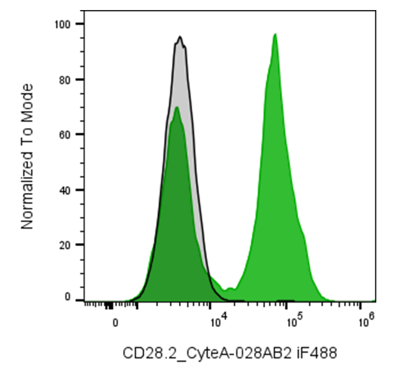
| Cat # | Size | Price | Quantity | |
|---|---|---|---|---|
| 180403 | 25 tests | $125 | ||
| 180404 | 100 tests | $250 |
| Clone | 028CB |
|---|---|
| Application | Flow Cytometry |
| Reactivity | Human |
| Format | iF488 |
| Target Name | CD28, Tp44 |
| Isotype | Rabbit IgG |
| Antibody Type | Monoclonal |
| Regulatory Status | RUO |
| Formulation | Phosphate-buffered solution, pH 7.2, containing 0.09% sodium azide and 0.2% (w/v) BSA |
| Protein Concentration | Supplied at a lot-specific concentration. |
| Storage & Handling | The antibody solution should be stored undiluted between 2°C and 8°C, and protected from prolonged exposure to light. Do not freeze. |
| Recommended Usage | For flow cytometric staining, it is recommended to use 5 µL of this reagent per 0.5-1.0 million cells in a 100 µL volume. Optimal reagent performance should be determined by titration for each specific application. iF488 has an excitation max at 491 nm and an emission max at 516 nm. |
| Excitation Laser | Blue Laser (488 nm) |
| Brand | InnoSite |
| See All Formats | Clone 028CB |
CD28 (Cluster of Differentiation 28) is a key costimulatory receptor expressed predominantly on T lymphocytes. It plays a vital role in the activation, proliferation, and survival of T-cells, complementing the antigen-specific signal delivered through the T-cell receptor (TCR). The engagement of CD28 is essential for full T-cell activation and the development of adaptive immune responses.
CD28 is a type I transmembrane glycoprotein belonging to the immunoglobulin (Ig) superfamily. Structurally, it exists as a homodimer on the T-cell surface and comprises three main parts: an extracellular Ig-like domain responsible for ligand binding, a transmembrane region, and a cytoplasmic tail containing signaling motifs. The intracellular region includes a YMNM motif that recruits PI3K (phosphoinositide 3-kinase) and adaptors such as Grb2 and GADS, promoting downstream signaling cascades including activation of Akt and NF-κB pathways. CD28 interacts primarily with B7 family ligands CD80 (B7-1) and CD86 (B7-2), expressed on professional antigen-presenting cells (APCs) such as dendritic cells, macrophages, and B cells. The binding of CD28 to these ligands, in conjunction with TCR engagement, enhances cytokine production (notably interleukin-2), cell metabolism, and survival, processes crucial for mounting effective immune responses.
Aberrant CD28 signaling is implicated in several pathological conditions. Overactivation can contribute to autoimmune diseases like rheumatoid arthritis and multiple sclerosis, where excessive T-cell stimulation leads to tissue damage. Conversely, reduced CD28 expression or signaling with aging (immunosenescence) can impair immune responses, leading to increased infection risk. Certain malignancies also exploit CD28 pathways to modulate immune evasion.
CD28 has been a therapeutic target and tool in immunotherapy. The development of CTLA-4-Ig fusion proteins (such as abatacept and belatacept) competitively blocks CD28-B7 interactions, reducing T-cell activation in autoimmune disorders and preventing transplant rejection. Conversely, engineered chimeric antigen receptor (CAR) T-cells often include CD28 intracellular domains to amplify antitumor activity by strengthening T-cell signaling. However, uncontrolled CD28 activation has proven hazardous, as exemplified by the TGN1412 trial, underscoring the need for careful modulation in therapeutic settings.
InnoSite™ iF488 Rabbit IgG, κ2 Isotype Control
InnoSite™ iF488 Anti-human CD28 Antibody TDS
Have a product or application question? Consult our FAQs or contact us.