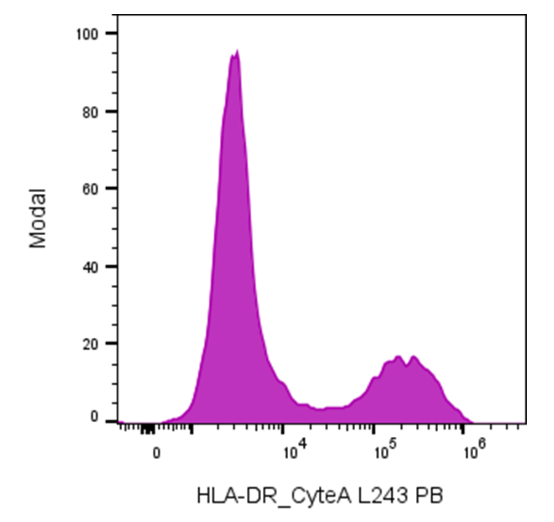
| Cat # | Size | Price | Quantity | |
|---|---|---|---|---|
| 180703 | 25 tests | $125 | ||
| 180704 | 100 tests | $250 |
| Clone | HLADRCB |
|---|---|
| Application | Flow Cytometry |
| Reactivity | Human |
| Format | PB |
| Target Name | HLA-DR, Major Histocompatibility Class II, MHC class II |
| Isotype | Rabbit IgG |
| Antibody Type | Monoclonal |
| Regulatory Status | RUO |
| Formulation | Phosphate-buffered solution, pH 7.2, containing 0.09% sodium azide and 0.2% (w/v) BSA |
| Protein Concentration | Supplied at a lot-specific concentration. |
| Storage & Handling | The antibody solution should be stored undiluted between 2°C and 8°C, and protected from prolonged exposure to light. Do not freeze. |
| Recommended Usage | For flow cytometric staining, it is recommended to use 5 µL of this reagent per 0.5-1.0 million cells in a 100 µL volume. Optimal reagent performance should be determined by titration for each specific application. Pacific Blue has an excitation max at 404 nm and an emission max at 455 nm. |
| Excitation Laser | Violet laser (405nm) |
| Brand | InnoSite |
| See All Formats | Clone HLADRCB |
HLA-DR is a major histocompatibility complex (MHC) class II molecule that plays a central role in adaptive immune responses by presenting antigenic peptides to CD4⁺ T helper cells. It is primarily expressed on professional antigen-presenting cells (APCs), including dendritic cells, macrophages, B cells, and thymic epithelial cells, and its expression can be induced on other cell types under inflammatory conditions, particularly by interferon-γ.
Structurally, HLA-DR is a heterodimer composed of an α chain (DRA) and a β chain (DRB), each containing two extracellular domains, a transmembrane region, and a short cytoplasmic tail. The α1 and β1 domains together form the peptide-binding groove, which accommodates peptides typically 13–25 amino acids in length. This groove is open at both ends, allowing for flexibility in peptide size. HLA-DR is highly polymorphic, particularly in the DRB genes, enabling the immune system to present a broad repertoire of antigenic peptides derived from pathogens or self-proteins. The ligands of HLA-DR are processed peptide antigens generated from extracellular or vesicular proteins that are internalized, degraded in endosomal compartments, and loaded onto HLA-DR molecules. Peptide loading is tightly regulated by accessory molecules, including the invariant chain (Ii), which prevents premature peptide binding, and HLA-DM, which facilitates peptide exchange and stabilizes high-affinity peptide–HLA-DR complexes. The primary functional interaction of HLA-DR is with the T cell receptor (TCR) on CD4⁺ T cells, initiating T cell activation and differentiation.
HLA-DR is strongly implicated in disease. Specific HLA-DR alleles are associated with susceptibility or protection in numerous autoimmune diseases, including rheumatoid arthritis, type 1 diabetes, systemic lupus erythematosus, and multiple sclerosis, reflecting differences in self-antigen presentation. Aberrant or reduced HLA-DR expression is also observed in cancer and sepsis, where impaired antigen presentation contributes to immune evasion or immunosuppression. Conversely, elevated HLA-DR expression on monocytes is often used as a marker of immune activation and immune competence.
Therapeutically, HLA-DR has both direct and indirect relevance. Anti-HLA-DR monoclonal antibodies have been explored in transplantation and hematologic malignancies to modulate immune responses or deplete malignant APCs. In cancer immunotherapy and vaccine development, effective antigen presentation via HLA-DR is essential for robust CD4⁺ T cell help, supporting durable antitumor and antiviral immunity. Additionally, HLA-DR expression is widely used as a diagnostic and prognostic biomarker in immunology, oncology, and critical care settings.
InnoSite™ PB Rabbit IgG, κ2 Isotype Control
InnoSite™ PB Anti-human HLA-DR Antibody TDS
Have a product or application question? Consult our FAQs or contact us.