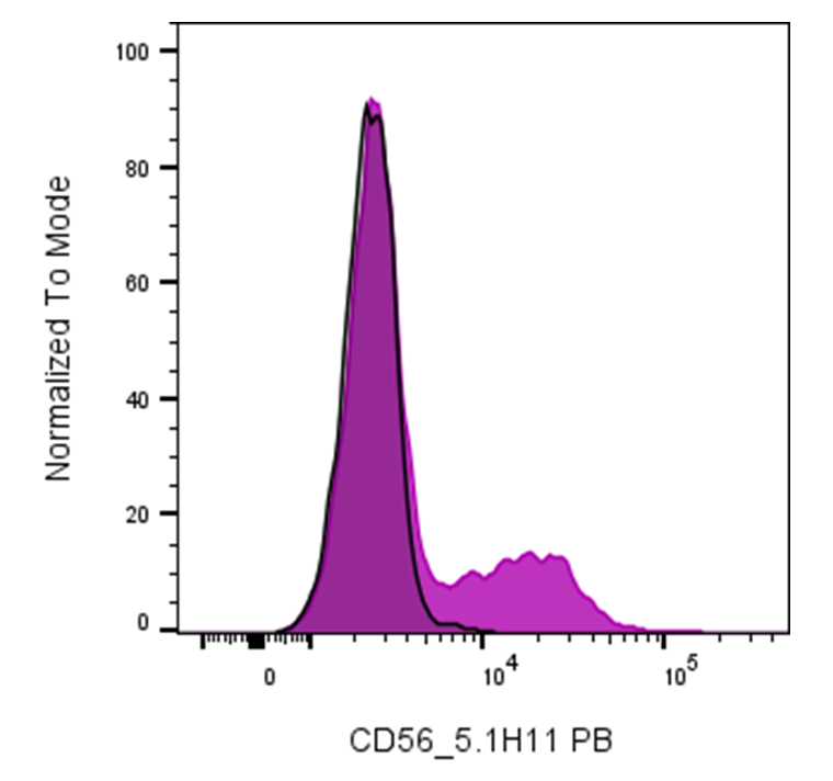
| Cat # | Size | Price | Quantity | |
|---|---|---|---|---|
| 115110 | 25 tests | $110 | ||
| 115111 | 100 tests | $240 |
| Clone | 5.1H11 |
|---|---|
| Application | Flow Cytometry |
| Reactivity | Human |
| Format | PB |
| Target Name | CD56, Leu-19, NKH1 |
| Isotype | mouse IgG1 |
| Antibody Type | Monoclonal |
| Regulatory Status | RUO |
| Formulation | Phosphate-buffered solution, pH 7.2, containing 0.09% sodium azide and 0.2% (w/v) BSA |
| Protein Concentration | Supplied at a lot-specific concentration. |
| Storage & Handling | The antibody solution should be stored undiluted between 2°C and 8°C, and protected from prolonged exposure to light. Do not freeze. |
| Recommended Usage | For flow cytometric staining, it is recommended to use 5 µL of this reagent per 0.5-1.0 million cells in a 100 µL volume. Optimal reagent performance should be determined by titration for each specific application. Pacific Blue has an excitation max at 404 nm and an emission max at 455 nm. |
| Excitation Laser | Violet laser (405nm) |
| See All Formats | Clone 5.1H11 |
CD56, also known as neural cell adhesion molecule (NCAM), is a multifunctional cell surface glycoprotein belonging to the immunoglobulin (Ig) superfamily. While originally characterized in the nervous system, CD56 is best known in immunology as a defining marker of natural killer (NK) cells and a subset of T lymphocytes. Its expression level on NK cells is used to distinguish functionally distinct subsets, most notably CD56bright and CD56dim NK cells.
Structurally, CD56 is a type I transmembrane protein composed of five extracellular Ig-like domains and two fibronectin type III domains, followed by a single transmembrane region and a cytoplasmic tail. Alternative splicing generates several isoforms (primarily NCAM-120, NCAM-140, and NCAM-180), which differ in their cytoplasmic domains and signaling capabilities. CD56 can also be post-translationally modified by polysialylation, a feature that modulates its adhesive properties and is particularly important in neural development.
Functionally, CD56 mediates homophilic interactions (CD56-CD56 binding) as well as heterophilic interactions with other ligands, including heparan sulfate proteoglycans, fibroblast growth factor receptor (FGFR), and components of the extracellular matrix. In the immune system, CD56 plays a role in cell-cell adhesion, immune synapse formation, and signal transduction. CD56bright NK cells are typically less cytotoxic but produce high levels of cytokines such as IFN-γ, contributing to immune regulation, whereas CD56dim NK cells exhibit potent cytotoxic activity against virally infected and transformed cells.
CD56 is implicated in several diseases. Aberrant or ectopic expression of CD56 is observed in various malignancies, including multiple myeloma, small cell lung carcinoma, neuroendocrine tumors, and certain leukemias and lymphomas. In hematologic diagnostics, CD56 expression is routinely used as an immunophenotypic marker to aid in disease classification and prognosis. For example, CD56 expression in multiple myeloma has been associated with altered patterns of bone marrow adhesion and disease behavior.
Therapeutically, CD56 has emerged as a potential target in oncology. Antibody-based approaches and antibody–drug conjugates directed against CD56 have been explored to selectively eliminate CD56-expressing tumor cells, particularly in neuroendocrine cancers and hematologic malignancies. In addition, CD56 is widely used as a marker for NK cell isolation, monitoring, and quality control in NK cell–based immunotherapies, underscoring its importance in both basic research and clinical applications.
PB Mouse IgG1 Isotype Control Antibody
PB Anti-Human CD56 Antibody TDS
Have a product or application question? Consult our FAQs or contact us.