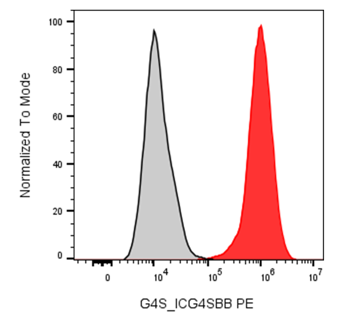
| Cat # | Size | Price | Quantity | |
|---|---|---|---|---|
| 360903 | 25 tests | $195 | ||
| 360904 | 100 tests | $460 |
| Clone | ICG4SBB |
|---|---|
| Application | Flow Cytometry |
| Reactivity | G4S Linker |
| Format | PE |
| Target Name | G4S Linker, GGGGS Linker, CAR linker |
| Isotype | Rabbit IgG |
| Antibody Type | Monoclonal |
| Regulatory Status | RUO |
| Formulation | Phosphate-buffered solution, pH 7.2, containing 0.09% sodium azide and 0.2% (w/v) BSA |
| Protein Concentration | Supplied at a lot-specific concentration. |
| Storage & Handling | The antibody solution should be stored undiluted between 2°C and 8°C, and protected from prolonged exposure to light. Do not freeze. |
| Recommended Usage | For flow cytometric staining, it is recommended to use 5 μL of this reagent per 0.5-1.0 million cells in a 100 µL volume. Optimal reagent performance should be determined by titration for each specific application. |
| Excitation Laser | Blue Laser (488 nm) Green/Yellow laser (532/561nm) |
| Research Areas | CAR, CAR-T, Chimeric Antigen Receptor, Immunotherapy |
| See All Formats | Clone ICG4SBB |
Anti-G4S linker antibody is an antibody designed to recognize the glycine-serine-rich G4S linker commonly incorporated into single-chain variable fragments (scFvs) and chimeric antigen receptors (CARs). The core sequence is Gly-Gly-Gly-Gly-Ser (GGGGS), which is frequently repeated three or four times to provide flexibility between the VH and VL domains of an scFv. Because the G4S sequence is largely independent of the antigen-binding specificity of the CAR, anti-G4S antibodies can serve as broadly applicable reagents for detecting different CAR constructs.
Structurally, the G4S linker is a flexible, hydrophilic peptide rather than a naturally occurring cell-surface protein. In an scFv-based CAR, it connects the variable heavy and variable light domains and helps maintain appropriate folding and flexibility of the antigen-binding domain. Anti-G4S antibodies recognize epitopes formed by repeated G4S sequences. Some validated monoclonal antibodies require two or more G4S repeats for optimal recognition.
The G4S linker has no physiological ligand or receptor because it is an engineered sequence. Consequently, anti-G4S antibodies do not have a conventional disease-associated target. Their principal role is analytical: they can detect CAR-positive cells independently of the CAR's target antigen, making them useful for flow cytometry, CAR-T cell characterization, monitoring, and manufacturing quality control. Recent studies have demonstrated sensitive detection across CARs directed against different antigens, including CD19 and CLDN6.
Anti-G4S antibodies may therefore support CAR-T therapy development rather than directly treating disease. They can help quantify CAR expression, assess transduction and cell-manufacturing quality, and monitor CAR-T cells during research or clinical studies. Because the same reagent can recognize multiple G4S-containing CAR designs, anti-G4S detection provides a potentially standardized, antigen-independent approach for CAR-T cell analysis and quality control.
APC Rabbit IgG, κ2 Isotype Control (Fc silent) Antibody
PE Anti-G4S Linker Antibody TDS
Have a product or application question? Consult our FAQs or contact us.