The antibody solution should be stored undiluted between 2°C and 8°C, and protected from prolonged exposure to light. Do not freeze.
Recommended Usage
For flow cytometric staining, it is recommended to use 5 uL of this reagent per 0.5-1.0 million cells in a 100 µL volume. Optimal reagent performance should be determined by titration for each specific application.
Excitation Laser
Blue Laser (488 nm), Green Laser (532 nm)/Yellow-Green Laser (561 nm)
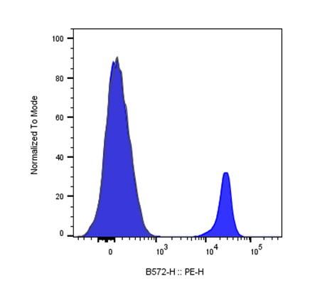
CD27 is a 50–55 kDa type I transmembrane glycoprotein, also known as S152 and T14, and is a lymphocyte-specific member of the tumor necrosis factor receptor (TNFR) superfamily, which also includes CD30 and CD40. Members of this superfamily are broadly involved in the regulation of cell proliferation, differentiation, and apoptosis. Unlike many other TNFR/TNF family members, expression of CD27 and its ligand CD70 is predominantly confined to lymphocytes, reflecting their specialized role in the adaptive immune response.
How do I determine the optimal antibody concentration/titer for my experiment?
We recommend titrating each new antibody lot on your own cell type and instrument, since optimal concentration depends on cell density, target expression level, and cytometer sensitivity. Suggested starting dilutions or test sizes (e.g., µg or µl per 10^6 cells) are provided on the product page as a starting reference point, not a fixed requirement.
Can these antibodies be used for intracellular or intranuclear staining, or only surface staining?
Intended use (surface, intracellular, or both) is specified per product, indicated under Application. The appropriate Related Protocol is linked for each product. If you need to detect both surface and intracellular markers in the same panel, confirm each antibody's compatibility with your fixation/permeabilization protocol before combining them.
What controls should I use alongside these antibodies?
Matched isotype controls (same host species, isotype, and conjugate) are available for most clones to help distinguish specific binding from background/non-specific staining and are linked on each product page. Unstained, single-color compensation, and fluorescence-minus-one (FMO) controls are also recommended, especially for multicolor panel design.
Can I combine InnoCyto Flow Cytometry antibodies into a multicolor panel?
Yes — antibodies across our catalog are designed to be panel-compatible, and conjugate options are offered specifically to support multiplexing. When building a panel, pair bright fluorophores with low-expression targets, avoid excessive spectral overlap, and confirm compensation is set up correctly for your specific fluorochrome combination. Use our Panel Builder to simplify the process.
Have a product or application question? Consult our FAQs or contact us.