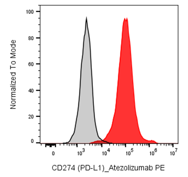
| Cat # | Size | Price | Quantity | |
|---|---|---|---|---|
| 119903 | 25 tests | $95 | ||
| 119904 | 100 tests | $215 |
| Clone | Atezolizumab |
|---|---|
| Application | Flow Cytometry |
| Reactivity | Human |
| Format | PE |
| Target Name | CD274, PD-L1, B7-H1, Programmed cell death ligand 1, B7 homolog 1 |
| Isotype | Human IgG1 |
| Antibody Type | Monoclonal |
| Regulatory Status | RUO |
| Formulation | Phosphate-buffered solution, pH 7.2, containing 0.09% sodium azide and 0.2% (w/v) BSA |
| Protein Concentration | Supplied at a lot-specific concentration. |
| Storage & Handling | The antibody solution should be stored undiluted between 2°C and 8°C, and protected from prolonged exposure to light. Do not freeze. |
| Recommended Usage | For flow cytometric staining, it is recommended to use 5 µL of this reagent per 0.5-1.0 million cells in a 100 µL volume. Optimal reagent performance should be determined by titration for each specific application. iF488 has an excitation max at 491 nm and an emission max at 516 nm. |
| Excitation Laser | Blue Laser (488 nm) Green/Yellow laser (532/561nm) |
| See All Formats | Clone Atezolizumab |
Human programmed death-ligand 1 (PD-L1), also known as CD274 or B7-H1, is an immune checkpoint protein that plays a central role in regulating T-cell activity and maintaining peripheral immune tolerance. PD-L1 is expressed on antigen-presenting cells, including dendritic cells and macrophages, and can be induced on many other cell types by inflammatory cytokines such as interferon-γ. It is frequently upregulated by tumor cells as a mechanism of immune evasion.
PD-L1 is a type I transmembrane glycoprotein of approximately 290 amino acids. Its extracellular region contains an N-terminal immunoglobulin variable (IgV)-like domain and a membrane-proximal IgC2-like domain, followed by a single transmembrane helix and a short cytoplasmic tail of approximately 30 amino acids. The IgV domain contains the primary binding interface for PD-1. Structural studies show that PD-L1 engages PD-1 through an extensive protein-protein interface involving hydrophobic and polar interactions.
The principal receptor for PD-L1 is programmed cell death protein 1 (PD-1/CD279), expressed mainly on activated T cells and other immune cells. PD-L1 can also interact with B7-1 (CD80), including interactions occurring in cis on antigen-presenting cells. Engagement of PD-1 by PD-L1 suppresses T-cell receptor signaling, proliferation, cytokine production, and cytotoxic activity, thereby limiting excessive immune responses.
Dysregulated PD-L1 expression contributes to immune suppression in cancer and has been associated with chronic infections and inflammatory diseases. Therapeutically, the PD-1/PD-L1 pathway is one of the most important targets in cancer immunotherapy. Monoclonal antibodies blocking PD-1 or PD-L1 can restore antitumor T-cell activity and have demonstrated substantial clinical benefit across multiple cancers. PD-L1 is therefore both a therapeutic target and an important biomarker for patient selection in certain immunotherapy settings.
PE Human IgG1 Isotype Control Antibody
PE Anti-Human CD274 (PD-L1) Antibody TDS
Have a product or application question? Consult our FAQs or contact us.