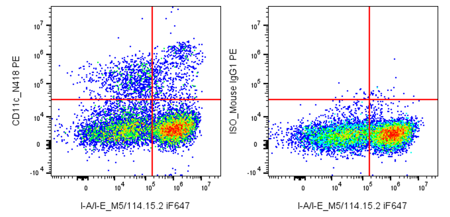
| Cat # | Size | Price | Quantity | |
|---|---|---|---|---|
| 201609 | 25 µg | $45 | ||
| 201610 | 100 µg | $105 |
| Clone | N418 |
|---|---|
| Application | Flow Cytometry |
| Reactivity | Mouse |
| Format | PE |
| Target Name | CD11c, integrin αX chain, CR4, ITGAX, p150 |
| Isotype | Armenian Hamster IgG |
| Antibody Type | Monoclonal |
| Regulatory Status | RUO |
| Formulation | Phosphate-buffered solution, pH 7.2, containing 0.09% sodium azide and 0.2% (w/v) BSA |
| Protein Concentration | 0.2 mg/mL |
| Storage & Handling | The antibody solution should be stored undiluted between 2°C and 8°C, and protected from prolonged exposure to light. Do not freeze. |
| Recommended Usage | For flow cytometric staining, it is recommended to use less than 0.5 µg of this reagent per 0.5-1.0 million cells in a 100 µL volume. Optimal reagent performance should be determined by titration for each specific application. PE has an excitation max at 565 nm and an emission max at 575 nm. |
| Excitation Laser | Blue Laser (488 nm) Green/Yellow laser (532/561nm) |
| See All Formats | Clone N418 |
CD11c, also known as integrin αX (ITGAX), is a cell surface adhesion and signaling molecule best known as a hallmark marker of dendritic cells in humans and mice. It is also expressed on subsets of monocytes, macrophages, natural killer (NK) cells, and activated T cells. CD11c plays an important role in immune surveillance by regulating cell adhesion, migration, and interactions between antigen-presenting cells and other immune cells.
Structurally, CD11c is a type I transmembrane glycoprotein that heterodimerizes with the integrin β2 subunit (CD18) to form the αXβ2 integrin, also known as complement receptor 4 (CR4). The extracellular domain of CD11c contains an inserted (I) domain, also referred to as an A domain, which is responsible for ligand binding and requires divalent cations such as Mg²⁺ or Mn²⁺ for activity. Like other integrins, CD11c undergoes conformational changes that regulate its affinity for ligands and enable bidirectional signaling. Its short cytoplasmic tail interacts with cytoskeletal and signaling adaptor proteins but lacks intrinsic enzymatic activity.
The primary ligands of CD11c include the complement fragment iC3b, fibrinogen, heparin, and several extracellular matrix proteins. Through binding to iC3b, CD11c contributes to complement-mediated phagocytosis and clearance of opsonized pathogens and apoptotic cells. CD11c also facilitates cell adhesion and migration across endothelial barriers, supporting the trafficking of dendritic cells and monocytes to sites of infection or inflammation and to secondary lymphoid organs.
CD11c is implicated in a range of diseases involving immune dysregulation. In inflammatory and autoimmune disorders, CD11c⁺ myeloid cells contribute to tissue inflammation and antigen presentation, sometimes exacerbating pathology. In cancer, CD11c⁺ dendritic cells play dual roles: they can support antitumor immunity by presenting tumor antigens to T cells, but dysfunctional or tolerogenic CD11c⁺ populations within the tumor microenvironment may promote immune evasion. Altered CD11c expression is also observed in chronic infections, where it can reflect changes in myeloid cell differentiation and function.
Therapeutically, CD11c is most commonly leveraged as a biomarker and targeting handle rather than a direct drug target. Antibodies against CD11c are widely used for the identification, isolation, and depletion of dendritic cells in research and preclinical models. In immunotherapy, CD11c⁺ dendritic cells are central to vaccine strategies, where they are loaded with tumor or pathogen-derived antigens to elicit robust T cell responses. Additionally, targeted delivery of antigens or immunomodulatory agents to CD11c⁺ cells is being explored to enhance vaccine efficacy and modulate immune responses in cancer and autoimmune disease.
PE Armenian Hamster IgG Isotype Ctrl
PE Anti-Mouse CD11c Antibody TDS
Have a product or application question? Consult our FAQs or contact us.