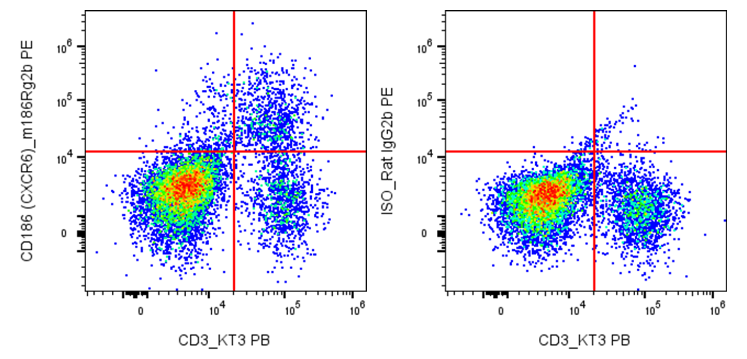
| Cat # | Size | Price | Quantity | |
|---|---|---|---|---|
| 205605 | 25 µg | $75 | ||
| 205606 | 100 µg | $160 |
| Clone | m186Rg2b |
|---|---|
| Application | Flow Cytometry |
| Reactivity | Mouse |
| Format | PE |
| Target Name | CD186, CXCR6, Chemokine (C-X-C motif) receptor 6 |
| Isotype | Rat IgG2b |
| Antibody Type | Monoclonal |
| Regulatory Status | RUO |
| Formulation | Phosphate-buffered solution, pH 7.2, containing 0.09% sodium azide and 0.2% (w/v) BSA |
| Protein Concentration | 0.2 mg/mL |
| Storage & Handling | The antibody solution should be stored undiluted between 2°C and 8°C, and protected from prolonged exposure to light. Do not freeze. |
| Recommended Usage | For flow cytometric staining, it is recommended to use less than 0.2 µg of this reagent per 0.5-1.0 million cells in a 100 µL volume. Optimal reagent performance should be determined by titration for each specific application. PE has an excitation max at 565 nm and an emission max at 575 nm. |
| Excitation Laser | Blue Laser (488 nm) Green/Yellow laser (532/561nm) |
| See All Formats | Clone m186Rg2b |
Mouse CD186, also known as C-X-C chemokine receptor 6 (CXCR6), is a seven-transmembrane G protein-coupled receptor (GPCR) encoded by the Cxcr6 gene. It is expressed on activated CD4+ and CD8+ T cells, natural killer (NK) cells, natural killer T (NKT) cells, subsets of γδ T cells, and tissue-resident memory T (TRM) cells. CXCR6 plays a critical role in directing immune cell migration, retention, and survival within peripheral tissues, particularly the liver, lungs, skin, and sites of chronic inflammation. It is especially important for establishing and maintaining tissue-resident lymphocyte populations that provide rapid local immune protection.
Structurally, mouse CD186 is a class A GPCR consisting of seven α-helical transmembrane domains connected by extracellular and intracellular loops, an extracellular N-terminus involved in ligand binding, and an intracellular C-terminal tail that interacts with heterotrimeric G proteins and signaling molecules. Its primary and essentially exclusive ligand is CXCL16, a unique chemokine that exists in both a soluble form, which functions as a chemoattractant, and a membrane-bound form, which also serves as an adhesion molecule. Binding of CXCL16 activates intracellular signaling pathways that promote chemotaxis, cell survival, adhesion, and cytokine production.
The CXCR6-CXCL16 axis contributes to numerous inflammatory and immune-mediated diseases. In mouse models, CXCR6 regulates lymphocyte recruitment during viral infections, autoimmune hepatitis, inflammatory bowel disease, asthma, and metabolic liver disease. Depending on the disease context, CXCR6-positive immune cells may either protect tissues by eliminating infected cells or promote pathology through excessive inflammation. In cancer, CXCR6-expressing CD8+ T cells and NK cells are frequently associated with improved antitumor immunity because they preferentially localize within tumors and exhibit enhanced cytotoxic function.
Because of its role in immune cell trafficking and tissue residency, CD186 has become an attractive therapeutic target. Inhibition of the CXCR6-CXCL16 pathway is being investigated to reduce chronic inflammatory diseases and transplant rejection, whereas strategies that enhance CXCR6-positive T-cell recruitment or persistence may improve cancer immunotherapy, including adoptive T-cell transfer and CAR-T cell therapies. Consequently, mouse CXCR6 is widely used to study tissue-resident immunity, chronic inflammation, infectious disease, and mechanisms of antitumor immune responses.
PE Anti-Mouse CD186 (CXCR6) Antibody TDS
Have a product or application question? Consult our FAQs or contact us.