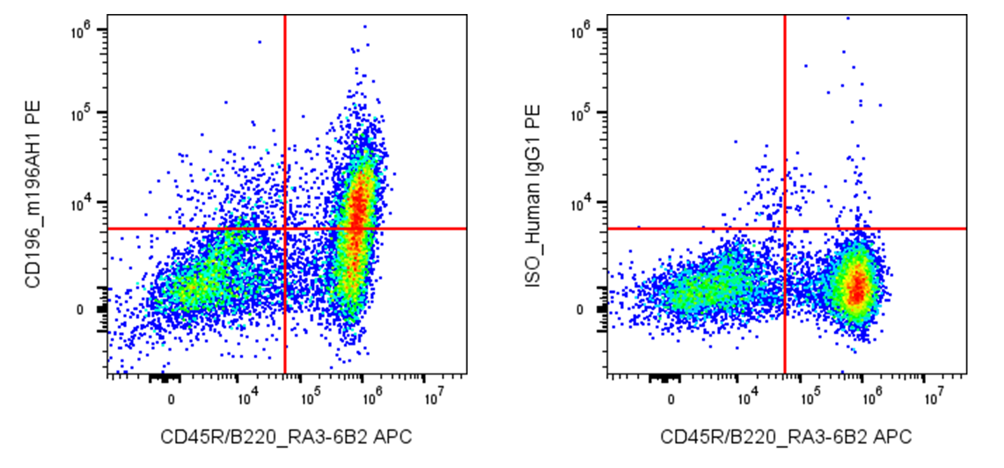
| Cat # | Size | Price | Quantity | |
|---|---|---|---|---|
| 208605 | 25 µg | $125 | ||
| 208606 | 100 µg | $295 |
| Clone | m196AH1 |
|---|---|
| Application | Flow Cytometry |
| Reactivity | Mouse |
| Format | PE |
| Target Name | CD196, CCR6, CD-196, DRY6, BN-1, CKR-L3, STRL22 |
| Isotype | Human IgG1 |
| Antibody Type | Monoclonal |
| Regulatory Status | RUO |
| Formulation | Phosphate-buffered solution, pH 7.2, containing 0.09% sodium azide and 0.2% (w/v) BSA |
| Protein Concentration | 0.2 mg/mL |
| Storage & Handling | The antibody solution should be stored undiluted between 2°C and 8°C, and protected from prolonged exposure to light. Do not freeze. |
| Recommended Usage | For flow cytometric staining, it is recommended to use less than 0.2 µg of this reagent per 0.5-1.0 million cells in a 100 µL volume. Optimal reagent performance should be determined by titration for each specific application. PE has an excitation max at 565 nm and an emission max at 575 nm. |
| Excitation Laser | Blue Laser (488 nm) Green/Yellow laser (532/561nm) |
| See All Formats | Clone m196AH1 |
CD196, also known as CCR6, is a chemokine receptor that regulates immune-cell migration, positioning, and inflammatory responses. It is expressed on several immune-cell populations, including memory and activated T cells, Th17 cells, regulatory T cells (Tregs), and subsets of B cells and dendritic cells. Through recognition of chemokines, CCR6 helps direct these cells to mucosal tissues and sites of inflammation, where they participate in immune surveillance, pathogen defense, and the regulation of immune responses.
CCR6 is a seven-transmembrane G protein-coupled receptor (GPCR) belonging to the class A chemokine receptor family. Its extracellular regions recognize chemokine ligands, while its intracellular domains couple to G proteins and activate signaling pathways that regulate chemotaxis, cell adhesion, and migration. The principal ligand of CCR6 is CCL20, also known as macrophage inflammatory protein-3 alpha (MIP-3α) or liver and activation-regulated chemokine (LARC). CCL20 is produced by epithelial cells and various immune and stromal cells, particularly during inflammation. The CCL20–CCR6 axis is especially important for recruiting CCR6-expressing cells to mucosal surfaces, including the intestinal tract and skin.
CCR6 contributes to immune defense at barrier tissues, but dysregulation of its signaling can promote chronic inflammation and autoimmune disease. The CCL20–CCR6 axis has been implicated in psoriasis, rheumatoid arthritis, inflammatory bowel disease, and multiple sclerosis, partly through the recruitment and positioning of Th17 cells and other inflammatory leukocytes. Its effects depend on the tissue and immune-cell population involved; CCR6 expression on regulatory T cells may also support immune suppression and tissue homeostasis. In cancer, this pathway can influence tumor-associated inflammation and immune-cell infiltration, with effects that vary by tumor type and microenvironment.
Therapeutically, CCR6 and CCL20 are being investigated as potential targets for controlling inflammatory and autoimmune diseases. Blocking this pathway may reduce the recruitment of pathogenic immune cells into affected tissues. Conversely, manipulating CCR6-mediated trafficking could help direct beneficial immune cells to specific tissues or tumors. Because CCR6 is expressed by both inflammatory and regulatory populations, therapeutic strategies must account for its context-dependent effects on immune protection, inflammation, and tumor immunity.
PE Human IgG1 Isotype Control Antibody
PE Anti-mouse CD196 (CCR6) Antibody TDS
Have a product or application question? Consult our FAQs or contact us.