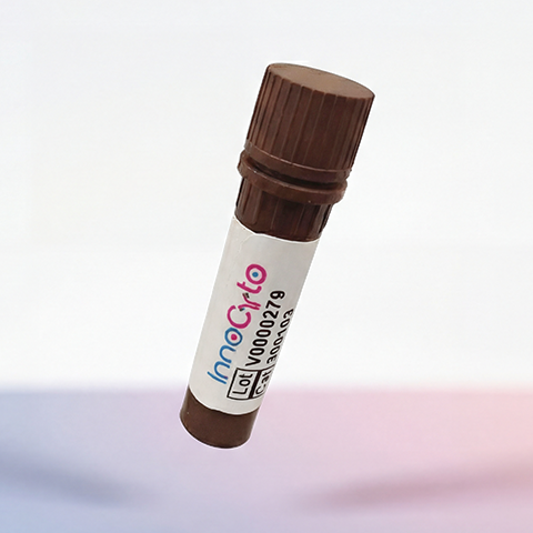
| Cat # | Size | Price | Quantity | |
|---|---|---|---|---|
| 803701 | 25 ug | $245 | ||
| 803702 | 100 ug | $595 |
| Application | Flow Cytometry |
|---|---|
| Format | Liquid, PE |
| Expression Host | CHO |
| Target Name | CD22, SIGLEC2, BL-CAM |
| Species | Human |
| Sources | Human CD22 protein (Asp20-Arg687) with C-terminus Fc tag is expressed in CHO cells and conjugated to PE. |
| Accession Number | P20273 |
| Molecular Weight | The protein has a predicted molecular weight of 101 kDa. Under DTT-reducing conditions, it migrates at approximately 130-150 kDa on SDS-PAGE prior to conjugation. |
| Affinity Tag | C-Fc |
| Regulatory Status | RUO |
| Formulation | 1xPBS buffer, pH7.4, 0.09% NaN3 with a carrier protein |
| Endotoxin level | Not tested |
| Protein Concentration | 25µg size is bottled at 0.1mg/mL concentration. 100 µg size is bottled at lot specific concentration. |
| Storage and Handling | Briefly centrifuge the vial upon receipt. An unopened vial may be stored at 2–8°C for up to six months. |
| Research Areas | B cells, Cancer Marker |
CD22 is a B cell–specific transmembrane glycoprotein that functions as an important regulator of B cell receptor (BCR) signaling and immune tolerance. Also known as Siglec-2, CD22 belongs to the sialic acid–binding immunoglobulin-like lectin (Siglec) family and is expressed almost exclusively on mature B cells, with expression increasing as B cells progress from the naïve to mature stages. Through its inhibitory signaling capacity, CD22 helps fine-tune B cell activation and prevent inappropriate immune responses.
Structurally, CD22 is a type I transmembrane protein with a large extracellular region composed of seven immunoglobulin-like domains. The N-terminal domain mediates binding to sialic acid–containing glycans, which serve as its primary ligands. CD22 preferentially recognizes α2,6-linked sialic acids that are commonly present on glycoproteins and glycolipids expressed on B cells themselves (cis interactions) as well as on neighboring cells (trans interactions). The cytoplasmic tail of CD22 contains multiple immunoreceptor tyrosine-based inhibitory motifs (ITIMs), which are essential for its signaling function. Functionally, CD22 acts as a negative regulator of BCR signaling. Upon BCR engagement, CD22 becomes phosphorylated and recruits phosphatases such as SHP-1 to its ITIM motifs. These phosphatases attenuate downstream signaling pathways, thereby raising the threshold for B cell activation. Through this mechanism, CD22 contributes to the maintenance of B cell tolerance and limits excessive antibody production. CD22 also influences B cell survival, migration, and interactions within lymphoid tissues.
Dysregulation of CD22 expression or signaling has been linked to immune-mediated diseases and malignancy. Reduced CD22 function can lead to hyperactive B cells and has been associated with autoimmune diseases such as systemic lupus erythematosus. In contrast, CD22 is frequently overexpressed on B cell malignancies, including B cell acute lymphoblastic leukemia (B-ALL) and certain non-Hodgkin lymphomas, making it an attractive diagnostic and therapeutic target.
CD22 plays a significant role in therapeutics, particularly in the treatment of B cell cancers. Antibody-based therapies targeting CD22 have been developed to selectively eliminate malignant B cells. Notably, antibody–drug conjugates and immunotoxins that bind CD22 deliver cytotoxic agents directly to cancerous B cells, sparing most non–B cell populations. CD22 is also being explored as a target for engineered cell therapies and for strategies aimed at modulating B cell activity in autoimmune disease. Together, these approaches highlight CD22 as a key molecule at the intersection of B cell biology, disease, and targeted therapy.
PE Human CD22 Protein (C-Fc) TDS
Anti-Human CD19 Antibody, Clone HIB19
iF560 Anti-Human CD19 Antibody, Clone HIB19
iF647 Anti-Human CD19 Antibody, Clone 019AM2b
Biotin Anti-Human CD19 Antibody, Clone 019AM2b
Anti-Human CD22 (Epratuzumab Biosimilar), Clone Epratuzumab
Anti-Human CD22 Antibody, Clone HIB22
iF647 Anti-Human CD22 Antibody, Clone HIB22
Human CD22 Protein (C-His-Avi)
Biotin Human CD22 Protein (C-His-Avi)
Have a product or application question? Consult our FAQs or contact us.