| Cat # | Size | Price | Quantity | |
|---|---|---|---|---|
| 804401 | 25 ug | $245 | ||
| 804402 | 100 ug | $595 |
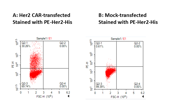
| Application | Flow Cytometry |
|---|---|
| Format | Liquid, PE |
| Expression Host | CHO |
| Target Name | HER2, HER-2, ERBB2, CD340, neu, MLN19, NEU, NGL, TKR1 |
| Species | Human |
| Sources | Recombinant Human Her2 protein (Thr23-Thr652) with C-terminus His tag is expressed in CHO cells and conjugated to PE. |
| Accession Number | P04626 |
| Molecular Weight | The protein has a predicted molecular weight of 71 kDa. Under DTT-reducing conditions, it migrates at approximately 85 kDa on SDS-PAGE prior to conjugation. |
| Affinity Tag | C-His |
| Regulatory Status | RUO |
| Formulation | 1xPBS buffer, pH7.4, 0.09% NaN3 with a carrier protein |
| Endotoxin level | Not tested |
| Protein Concentration | 25µg size is bottled at 0.1mg/mL concentration. 100 µg size is bottled at lot specific concentration. |
| Storage and Handling | Briefly centrifuge the vial upon receipt. An unopened vial may be stored at 2–8°C for up to six months. |
| Research Areas | Mesenchymal Stem Cells, Embryogenesis, Cancer Marker, Cancer Immunology |
HER2 (Human Epidermal Growth Factor Receptor 2), also known as ERBB2 or CD340, is a transmembrane protein that plays a pivotal role in normal cell growth and differentiation. It is a member of the epidermal growth factor receptor (EGFR/ERBB) family of receptor tyrosine kinases. Under normal physiological conditions, HER2 is expressed at low levels on the surface of epithelial cells, where it helps regulate cell proliferation and survival signals. However, its primary fame in medicine comes from its potent ability to drive uncontrolled cell growth when the gene encoding it is amplified or the protein is overexpressed.
Structurally, HER2 consists of an extracellular ligand-binding domain, a transmembrane spanning region, and an intracellular tyrosine kinase domain. A unique and critical feature of HER2 is that it is an "orphan receptor," meaning it has no known direct ligand. Unlike other family members (EGFR, HER3, HER4) that require a growth factor to bind and activate them, HER2 exists in a constitutively open conformation, ready to interact. It functions by forming heterodimers with other ligand-bound members of the HER family. This makes HER2 the preferred dimerization partner for all other ERBB receptors, amplifying the signaling strength of the network significantly.
In the context of disease, HER2 is a major driver of tumorigenesis. Gene amplification leads to the overexpression of HER2 proteins on the cell surface, sometimes up to 100 times the normal level. This results in spontaneous dimerization and continuous, ligand-independent firing of growth signals, leading to aggressive cell division and resistance to apoptosis. HER2 overexpression is most notably observed in approximately 15-20% of breast cancers and a significant subset of gastric and gastroesophageal cancers, classifying them as "HER2-positive."
Therapeutically, HER2 is one of the most successful targets in the history of precision oncology. The development of trastuzumab (Herceptin), a monoclonal antibody that binds to the extracellular domain of HER2, revolutionized treatment by blocking downstream signaling and flagging cells for immune destruction. Therapy has since evolved to include dimerization inhibitors like pertuzumab, small-molecule tyrosine kinase inhibitors (TKIs) like lapatinib that work inside the cell, and antibody-drug conjugates (ADCs) like T-DM1 and trastuzumab deruxtecan. These ADCs use the HER2 antibody as a "Trojan horse" to deliver potent chemotherapy directly into the cancer cell, sparing healthy tissue.
PE Human HER2 (CD340) Protein (C-His) TDS
Anti-Human HER2 (Pertuzumab Biosimilar), Clone Pertuzumab
Anti-Human HER2 (Trastuzumab Biosimilar), Clone Trastuzumab
Anti-Human HER2 (Trastuzumab Biosimilar), Clone Trastuzumab
Human HER2 (CD340) Protein (C-His-Avi)
Biotin Human HER2 (CD340) Protein (C-His-Avi)
Human HER2 (CD340) Protein (C-His)
Have a product or application question? Consult our FAQs or contact us.